Images in internal medicine
Received: 07 July 2019
Accepted: 20 November 2019
A 50-year-old male consulted due to a five-year history of heartburn, with esophageal dysphagia over the last year. Esophagoscopy showed a foreign body consisting of plant material; after removing it, narrowing (< 12.5 mm) was found which blocked the endoscope's passage. Proximal to this narrowing were esophageal mucosal breaks spanning less than 75% of the circumference, with a maximum length of 1.5 cm. No biopsies were taken due to mucosal friability. (Figures 1, 2). The diagnosis was: esophageal stenosis secondary to gastroesophageal reflux (GER), suspected esophageal metaplasia (Barrett's esophagus) and grade C esophagitis. The current diagnostic model for GER revolves around the identifi cation of esophageal and extraesophageal symptoms, as well as mucosal damage (esophagitis, Barrett's esophagus, stenosis and adenocarcinoma) 1,2. This case illustrates three esophageal complications caused by GER. Esophageal dilation, treatment with omeprazole and follow up to histologically rule out Barrett's esophagus were ordered.
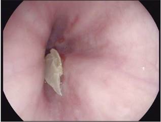
Figura 1
Cuerpo extraño esofágico constituído por material vegetal.
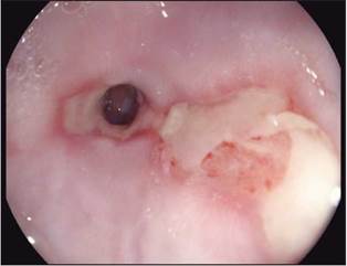
Figura 2
Estenosis esofágica, Esofagitis grado C y sospecha endoscópica de metaplasia esofágica.
References
1. Vakil N, van Zanten SV, Kahrilas P. Dent J, Jones R. The Montreal definition and classification of gastroesophageal reflux disease: a global evidence-based Consensus. Am J gastroneterol 2006; 101: 1900-20
2. Gyawali C, Kahrilas P , Savarino E, Zerbib F, Mion F, Somut A, et al. Modern diagnosis of GERD: the Lyon Consensus. Gut online first, published on February 9, 2018 as 10.1136/gutjnl 2017-314722
Notes
Author notes
* Correspondencia: Henry Alberto Royero Gutiérrez. Ocaña (Norte de Santander). E-mail: royerogastro@hotmail.com
