Images in Internal Medicine
Pulmonary alveolar microlithiasis. An incidental finding in a patient with COPD
Microlitiasis alveolar pulmonar. Un hallazgo incidental en un paciente con EPOC
Pulmonary alveolar microlithiasis. An incidental finding in a patient with COPD
Acta Medica Colombiana, vol. 46, no. 2, p. 51, 2021
Asociacion Colombiana de Medicina Interna
Received: 15 August 2020
Accepted: 27 October 2020
A 67-year-old male with a history of chronic obstructive pul monary disease and heavy smoking consulted due to progressive worsening of his dyspnea over the previous week along with a dry cough and unquantified fever at home. A chest x-ray showed bilateral calcified micronodules. A high-resolution computerized tomography confirmed these findings. The diagnosis was confirmed by the histological results of a bronchoalveolar lavage showing characteristic laminar microliths.
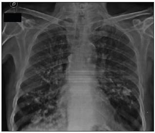
Figure
1. Chest x-ray showing a dense, bilateral micronodular pattern in the perihilar region.

Figure
2. Axial high-resolution computerized tomography of the chest: A. Mediastinal window showing multiple bilateral calci fied densitites in the lower lobes. B. Pulmonary window showing bilateral calcified interlobular micronodular hyperdensities.
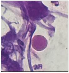
Figure
3. Hematoxylin and eosin staining of bronchoalveolar lavage liquid (100x) showing the presence of calcospherites
Pulmonary alveolar microlithiasis is a rare hereditary pul monary disease characterized by microcalcifications within the alveolar spaces 1. It is caused by mutations of the SCL34A2 gene which encodes the phosphate cotransporter in type II alveolar cells. This results in increased phosphate and calcium in lung surfactant, which leads to the formation and deposition of microliths within the alveoli as well as in other parts of the body 2. The definitive diagnosis requires histological identification of the microliths 3.
References
Menon PD, Hackman S. Pulmonary Alveolar Microlithiasis: An Isolated Case in a Hispanic Male. Case Rep Pathol. 2020:1-4.
Shaw BM, Shaw SD, McCormack FX. Pulmonary Alveolar Microlithiasis. Semin Respir Crit Care Med. 2020;41(2):280-7.
Tachibana T, Hagiwara K, Johkoh T. Pulmonary alveolar microlithiasis: Review and management. Curr Opin Pulm Med. 2009;15(5):486-90.
Author notes
*Correspondencia: Dr. Juan Pablo García-Marmolejo. Cali (Colombia). E-Mail: jpgarciamarmolejo@gmail.com