Ciências da Saúde
In vivo protective effect of cinnamon aqueous extract in carbon tetrachloride-treated male albino rats
In vivo protective effect of cinnamon aqueous extract in carbon tetrachloride-treated male albino rats
Acta Scientiarum. Health Sciences, vol. 43, e52826, 2021
Universidade Estadual de Maringá
Recepción: 27 Marzo 2020
Aprobación: 23 Abril 2021
Abstract: The liver as an organ is important for the metabolism of drugs and toxins. However, it is not immune from environmental insults. Exposure of liver cells to carbon tetrachloride (CCl.) results in the generation of tricholoromethyl radicals, which induce liver toxicity. This study aims at investigating the ameliorative effect of the cinnamon aqueous extract (CAE) against CCl.-induced hepatotoxicity in male albino rats. Hepatotoxicity was induced in rats through the intraperitoneal administration of 0.5 mL kg-1 body weight of CCl.. The analyses of the results obtained showed significant reduction in the levels of serum biochemical markers for 400 and 600 mg kg-1 bw of CAE protected rats as compared with CCl. group. In addition, CAE administration reversed liver tissue damaged via increased antioxidants markers. Histopathological examination of CAE treatment on rats showed improved changes to the liver damage caused by CCl. with no evidence of steatosis and inflammation. This result hence suggests that CAE has marked hepatoprotective and healing activities against CCl.-induced liver damage and could serve as a suitable candidate in drug discovery for the treatment of liver toxicity.
Keywords: Hepatotoxicity, Cinnamomum zeylanicum, carbon tetrachloride, liver damage.
Introduction
The liver, an important organ in the body that help sustain human life. It plays an important role to dominate numerous physiological processes which include different vital activities such as metabolism of drugs and toxins, secretion and storage (Shanmugasundaram & Venkataraman, 2006). Different stressors such as infectious agents, environmental pollutants, and hepatotoxins (carbon tetrachloride) are known to cause liver injuries (Kim et al., 2014). Carbon tetrachloride (CCl4) is classified as one of the most harmful substances which is usually employed for the induction of liver injuries in experimental rats (Kim et al., 2014). The exposure to CCl4 results in the generation of tricholoromethyl radicals which induce toxicity in rat liver (El-Sayed, Fouda, Mansour, & Elazab, 2015), raises the levels of hepatic lipid peroxidation and ultimately leading liver damage (Weber, Boll, & Stampfl, 2003). Reactive oxygen species in addition to the decrease in the body’s inhibitory and scavenging mechanism are responsible for its increased oxidative stress. For this, the liver cells have developed a defense mechanism to combat oxidative stress such as catalase, superoxide dismutase and glutathione peroxidase as well as antioxidants substances such as vitamin E, ascorbic and GSH (Kaplowitz & Tsukamoto, 1996).
When the orally ingested chemicals and drugs inter into the body system, they firstly go to the liver organ which represent the target part for the toxicity of these substances (xenobiotics and drugs), in which they are metabolized into a substances with intermediate toxicity. Therefor a huge numbers of xenobiotics and drugs are probably described as a hepatotoxic substances (Ajith, Hema, & Aswathy, 2007).
When the liver is exposed to different kinds of stress, it becomes damaged and it is thus incapable of performing its routine functions. Due to that, an urgent medical care is essentially needed when the liver is damaged. This damage occurs gradually and accumulates over many years which leads to liver disease (Garcia-Tsao & Lim, 2009). Traditional plants are common medications employed by most of the population (Garcia-Tsao & Lim, 2009; Ghashghaii, Hashemnia, Nikousefat, Zangeneh, & Zangeneh, 2017; Goorani et al., 2018). The impression that traditional plants and their role in prevention and treatment cannot be dispensed (Najafi et al., 2017; Zangeneh, Zangeneh, Tahvilian, & Moradi, 2018). Cinnamon (Cinnamomum zeylanicum) is a popular flavoring component, chiefly utilized in food manufacturing. Many studies have revealed that cinnamon has therapeutic and preventive effects against many diseases such as diabetes and glucose intolerance control (Khan, Safdar, Ali Khan, Khattak, & Anderson, 2003; Allen, Schwartzman, Baker, Coleman, & Phung, 2013), total cholesterol (Khan et al., 2003; Subash Babu, Prabuseenivasan, & Ignacimuthu, 2007), microbial diseases (Young & Oberg, 2000; Fabian, Sabol, Domaracka, & Bujnakova, 2006; Shahverdi, Monsef-Esfahani, Tavasoli, Zaheri, & Mirjani, 2007; Ranasinghe et al., 2013), anti-inflammatory (Yen, Lin, & Chang, 2010; Hong et al., 2012), the propagation of different cancer cell lines (Mancini-Filho, Van-Koiij, Mancini, Cozzolino, & Torres, 1998; Ka et al., 2003; Nishida et al., 2003), gastric lymphoma (Ozbayer et al., 2014) and antioxidant (Okawa, Kinjo, Nohara, & Ono, 2001; Murcia et al., 2004; Shen et al., 2012). Cinnamon extracts can affect β-carotene-linoleic acid through decreasing lipid peroxidation (Mancini-Filho et al., 1998). The effects of the antioxidant levels in cinnamon extract have previously being studied on carbon tetrachloride (CCl4)-induced hepatotoxicity in animal (Fu, Zheng, Lin, Ryerse, & Chen, 2008).
This study investigates the ameliorative effect of the cinnamon aqueous extract (CAE) on CCl4-induced hepatotoxicity in rats through the quantification of its potential antioxidant activities, the serum biochemical and histopathological analysis.
Material and methods
Plant materials
The barks of Cinnamon (Cinnamomum zeylanicum) were obtained from a local traditional market in Jeddah, Saudi Arabia and were identified by a Taxonomist. The barks were air dried at room temperature, washed with distilled water, dried again and grounded to fine powder. For the preparation of 10% decoction, 10 g of the Cinnamon bark powder was dissolved in 100 mL of distilled water and allowed to boil for about 30 minutes. The decoction was then cooled at room temperature, filtered and dispensed into a clean sterilized bottle; the bottles were then put into a refrigerator for storage until use.
Chromatographic analysis of Cinnamon extract (CE) by GC–MS
For the identification of bioactive compounds, GC–MS Chromatographic analysis of CE was performed using Agilent Technologies 7890B GC Systems equipped with 5977A Mass Selective Detector. Capillary column (HP-5MS Capillary; measuring 30.0m × 0.25 mm ID × 0.25 μm film) with helium as a carrier gas following a flow rate of 1.7 mL min.-1 and 1 μl injection. Analysis of the sample was carried out with the column held for a period of 4 min. at 40˚C post injection, followed by elevation in temperature to 300°C (20°C min.-1 heating ramp) along with a 3.0 min. hold. A split-less mode injection was applied at 300°C. MS scan range was (m z-1): 50 - 550 atomic mass units (AMU) under electron impact (EI) ionization (70 eV). The bioactive compounds were identified by comparing the retention indexes of these compounds together with their spectra mass with the NIST library in the GC-MS system.
Animals and treatment
Male Albino rats weighing 203.30 ± 0.93 g were bought from the animal house of Department of Biology, King Abdulaziz University, Jeddah and housed to acclimatized in the laboratory for one week. They were kept under normal laboratory temperature (23 ± 2ºC) with daily 12 hours of light on/off cycle, fed with standard feeds and body weight gained was recorded weekly. All the experimental studies relating to the animals were conducted following the National Institutes of Health Guide for the Care and Use of Laboratory Animals (NIH 1985); and in accordance with the World Medical Association Declaration of Helsinki regarding ethical conduct of research involving human subjects and/or animals.
The rats were separated into 8 groups with n = 6 has outlined below:
Group 1 (control): normal diet and water only.
Group 2 (CCl4): subcutaneous injection of 0.5 mL Kg-1 bw (20% CCl4 in paraffin oil) as previously described (Hesami et al., 2014) and commences from the start of the experiment.
Group 3 (CAE 400): 400 mg kg-1 bw of CAE orally for 4 weeks.
Group 4 (CCl4 + CAE400): oral protective dose of 400 mg kg -1 bw of CAE daily for 4 weeks + a single dose of 0.5 mL Kg-1 bw (20% CCl4 in paraffin oil) twice a day.
Group 5 (CCl4 + CAE600): oral protective dose of 600 mg kg-1 bw of CAE daily for 4 weeks + a single dose of 0.5 mL Kg-1 bw (20% CCl4 in paraffin oil) twice a day.
Group 6 (CCl4 + PCAE400): This post-treatment group was subcutaneously injected with 0.5 mL Kg-1 bw of CCl4 (20% CCl4 in paraffin oil) on the 14th day and then received oral dose of 400 mg kg bw CAE till experiment ends.
Group 7 (CCl4 + PCAE600): This post-treatment group was subcutaneously injected with 0.5 mL Kg-1 bw of CCl4 (20% CCl4 in paraffin oil) on the 14th day and then received oral dose of 600 mg . kg bw CAE till experiment ends
Group 8 (Silm): Oral dose of 100 mg kg-1 bw silymarin daily for 4 weeks (positive control).
Serum biochemical assay
At the conclusion of the experiment, the rats were anesthetized with pentobarbitone sodium (60 mg kg-1) after an overnight fast. Blood samples were dispensed in a centrifuge tube through cardiac puncture, kept to clot at 25°C for 15 minutes and centrifuged for 15 min. at 3000 rpm and 4°C. The serum levels of albumin (ALB) , alkaline phosphatase (ALP), HDL, Alanine Aminotransferase (Theocharis, Margeli, Skaltsas, Spiliopoulou, & Koutselinis, 2001), total protein (TP), total bilirubin (TBIL), Aspartate Aminotransferase (AST), LDL, triglyceride (TRIGL), Gamma-glutamyl transferase (GGT), cholesterol (Gäbele et al., 2009), glucose (GLUC), uric acid (UA) and urea were performed using a commercial assay kits and following to the manufacturer’s instruction (EGY-CHEM for lab technology, Bader city, Egypt).
Oxidative stress markers
The liver homogenate was done by homogenizing the liver tissue in 10 volumes of ice-cold 100 mM phosphate buffer containing 1 mM EDTA pH 7.4 using a homogenizer and centrifuged for 15 min at 10,000 rpm and 4°C. The resulting supernatant was utilized to determine the activities of liver enzymes markers of oxidative stress (SOD, GST, CAT) and Malondialdehyde (MDA) levels of lipid peroxidation. These assays were performed using commercial kits (Bioassay Technology Laboratory, Shanghai Korian Biotech CO).
Histopathological analysis
Liver samples were fixed in 10% neutral buffered formalin solution once sacrificed, routinely processed, paraffin embedded, sectioned at a thickness of 4 μm using a microtome, and hematoxylin and eosin (H&E) stained. All morphological alterations were examined using light microcopy.
Statistical analysis
Statistical analysis of all data was done with SPSS version 20 software. Test of significance was achieved by employing Student t-test. Significance level p was set at p < 0.05.
Results
GC-MS investigation of cinnamon extract (CE)
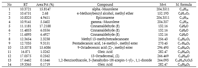
Analysis of CE using GC-MS showed the phytochemical components as shown in Figure 1 and Table 1. The GC-MS record described that the occurrences of many bioactive compounds which are arranged alongside with retention time, relative area (%), molecular weight and chemical formula (Table 1). By matching the spectra mass of the components against the NIST library, 14 major compounds peaks were identified from the 32 phytochemicals in CE. The major bioactive compound was Cinnamaldehyde (17.22%) followed by alpha-Muurolene (15.81%), 9-Octadecenoic acid (Z)-, methyl ester (15.61%), Pentadecanoic acid (9.31%), 14-methyl-, methyl ester and gamma.-Muurolene (5.54%).
Body weight
As illustrated in Table 2 below, a significant increase in body weight was observed in all experimental groups except in rats treated with CCl4, wherein a significant reduction in body weight (4.03 ± 1.39) was observed.
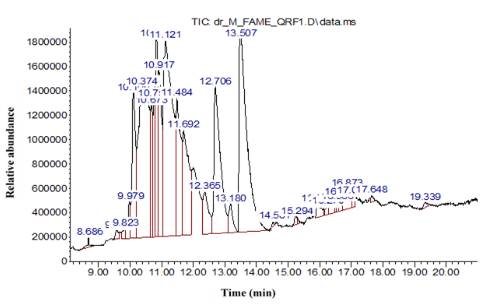
Figure 1. GCMS chromatogram of the n-hexane extract of Cinnamomum zeylanicum.
R.T. = retention time, Area Pct= peak area and Mwt= molecular weight.
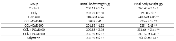
Data were stated as mean ± SEM and test of significance was achieved using Student t-test with significant differences at p< 0.05 and p< 0.01 as shown for (*) and (**) with respect to initial body weight.
Effects of CAE on serum biochemical parameters
The serum level of rats in the CCl4 treated group, showed a significant rise in the levels of ALP, ALT, AST, total bilirubin, cholesterol, GGT, glucose, LDL triglyceride, uric acid and urea whereas there were significant reduction in the HDL, TP and ALB levels in comparison with the control group (p < 0.001) (Table 3). However, rats pretreated with 400 and 600 mg kg-1 of CAE for four weeks as well as post-treatment with 400 and 600 mg kg-1 of CAE two weeks after the injection of CCl4, showed a significant reduction in the serum levels of ALT, AST, ALP, TC, TG, LDL, and glucose and a rise in the levels albumin and HDL (Table 3).
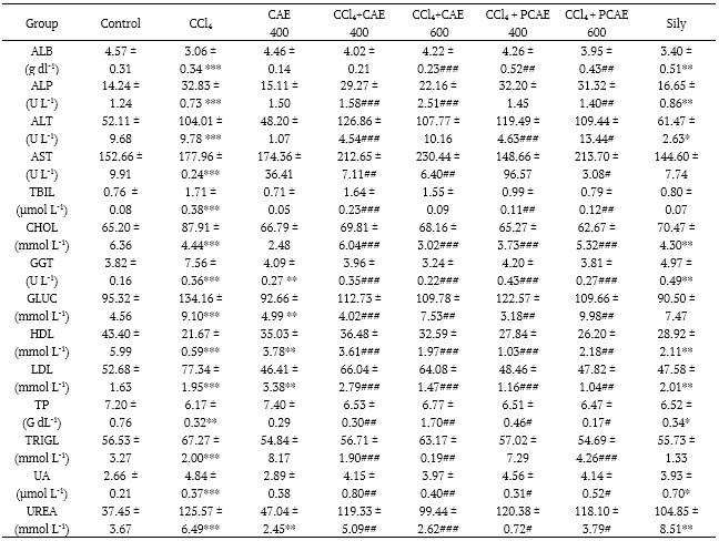
Data were stated as mean ± SEM and test of significance was achieved using Student t-test with significant differences at p < 0.01, p < 0.05, p < 0.001 as shown for (*),(**) and (***) with respect to normal control and by (#),(##),(###) with respect to CCl4 treated group respectively.
Effect of CAE on CCl4-induced oxidative stress
Animals treated with CCl4 revealed a significant reduction (p < 0.001) in the hepatic homogenate levels of SOD, CAT and GSH accompanied by significant increase in lipid peroxidation (p < 0.001) has quantified in the MDA levels in the hepatic tissue homogenate in comparison with the animals in the normal control group (Figure 2). Moreover, pretreatment with 400 and 600 gm kg-1 bw CAE resulted in a significant rise (p < 0.001) in hepatic tissue homogenate levels of SOD, CAT and GSH in comparison with CCl4 treated group. In addition, treatment with 400 and 600 gm kg-1 bw CAE significantly decreased (p < 0.001) the elevated hepatic tissue homogenate levels of MDA in comparison with CCl4 group (Figure 2).
Histopathological analysis
The micrographs of the histopathological analyses using haematoxilin-eosin staining is shown in (Figure 3 A-H).
The micrograph of rats in the normal control group as well as the CAE treated groups showed a normal aspect, healthy, cellular construction and clear nucleus within its cytoplasm (Figure 3A and 3C). However, exposure of liver tissue to CCl4 intoxication revealed deterioration in the feathery of the hepatic cells, steatosis, inflammation of the portal, sustained venous congestion and fibrosis (Figure 3B). Histopathological examination of liver tissue exposed to CCl4 as well as daily oral dose of 400 mg/kgbw of CAE showed evident sustained venous congestion with feathery deterioration, absence of steatosis or small vesicular steatosis, minimal parenchymal and portal inflammation (Figure 3D). In addition, findings on liver sample from animals in CCl4 group as well as daily oral dose of 600 mg kg-1 bw of CAE revealed an enhancement of the structural changes to the liver; with absence of sustained venous congestion, absence of steatosis, minimal parenchymal inflammation (Figure 3E).
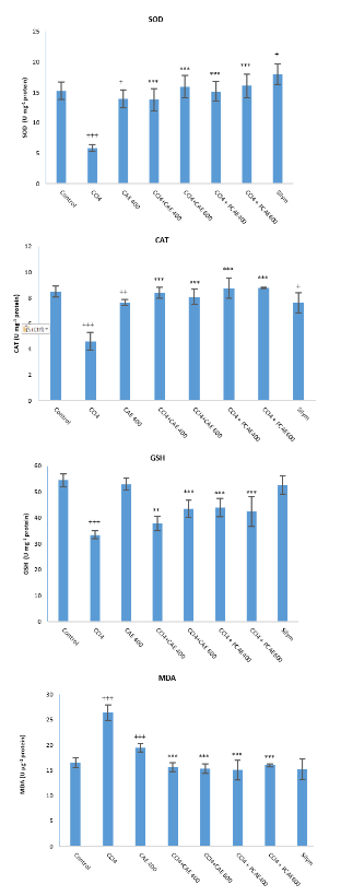
Figure 2. Effect of CAE on CCl4-induced oxidative stress. Data were stated as mean ± SEM and test of significance was achieved using Student t-test with significant differences at p < 0.05, p < 0.01 and p < 0.001 as shown for (+), (++) & (+++) in comparison with normal control and for (*), (**) & (***) in comparison with CCl4 group, respectively.
![Figure 3. Effect of CAE on histopathological changes of liver tissues (20x). (A) Control group showed typical histological structure of hepatic lobule with central vein [arrowhead] bordering typical hepatic cells [arrows]. (B) CCl4 exposed group showed sustained venous congestion and hepatocytes with feathery disintegration [arrows]. (C) CAE group (400 mg kg-1 bw) showed typical histological structure of hepatic lobule with central vein bordering typical hepatocytes [arrows]. (D) CCl4 + CAE group (400 mg kg-1 bw) showed evident sustained venous congestion with feathery disintegration, absence of steatosis, minimal portal and parenchymal inflammation [arrows]. (E) CCl4 + CAE group (600 mg kg-1 bw) showed hepatocytes structural enhancement with minimal vesicular steatosis [arrows]. (F) CCl4 + post-treatment of CAE group 400 mg/kg bw and (G) 600 mg kg-1 bw showed less percentages of necrosis regenerating hepatocytes. (H) Silymarin group showed hepatoprotective activity with normal sinusoids [arrow] and intact portal veins [arrowhead].](../307269997033_gf4.png)
Figure 3. Effect of CAE on histopathological changes of liver tissues (20x). (A) Control group showed typical histological structure of hepatic lobule with central vein [arrowhead] bordering typical hepatic cells [arrows]. (B) CCl4 exposed group showed sustained venous congestion and hepatocytes with feathery disintegration [arrows]. (C) CAE group (400 mg kg-1 bw) showed typical histological structure of hepatic lobule with central vein bordering typical hepatocytes [arrows]. (D) CCl4 + CAE group (400 mg kg-1 bw) showed evident sustained venous congestion with feathery disintegration, absence of steatosis, minimal portal and parenchymal inflammation [arrows]. (E) CCl4 + CAE group (600 mg kg-1 bw) showed hepatocytes structural enhancement with minimal vesicular steatosis [arrows]. (F) CCl4 + post-treatment of CAE group 400 mg/kg bw and (G) 600 mg kg-1 bw showed less percentages of necrosis regenerating hepatocytes. (H) Silymarin group showed hepatoprotective activity with normal sinusoids [arrow] and intact portal veins [arrowhead].
Discussion
The current study revealed the hepatoprotective, antioxidant and restorative effects of aqueous extract of cinnamon on CCl4-induced liver damage in rats. The liver as an important organ is responsible for drugs and xenobiotics metabolism. Various studies illustrated that CCl4 is commonly utilized in the induction of liver injury since cytochrome P450 is responsible for its metabolism in hepatic cells, producing a strongly reactive trichloromethyl radical, that initiates a sequential events of lipid peroxidation, cellular impairment and thus leading to liver fibrosis (Shenoy, Somayaji, & Bairy, 2001; Weber et al., 2003; Fang, Lai, & Lin, 2008; Halliwell & Gutteridge, 2015). Apart of lipid peroxidation, CCl4 is also a culprit in the depletion of tissue’s CAT, SOD and GSH actions, and this could be due to oxidative alteration of those proteins (Augustyniak, Waszkiewicz, & Skrzydlewska, 2005). This damage was related to loss of the liver function stated as a substantial rise in the total bilirubin content in the serum and reduction in albumin, which are proposed as markers of hepatic function (Renugadevi & Prabu, 2010).
The results of this study revealed that oral administration of aqueous extract of cinnamon efficiently protected against the harmful loss of those antioxidant actions after CCl4 injection, besides it is recognized to serve various biological functions, comprising cell protection from oxidative stress mediated by reactive oxygen species and free radicals (Nakamura, Torikai, & Ohigashi, 2001;Gäbele et al., 2009;). Bioactive compounds present in cinnamon were found to stimulate production of detoxifying agents and antioxidant enzymes in the transcriptional stage, through response elements of antioxidant (Masella, Di Benedetto, Varì, Filesi, & Giovannini, 2005) and to increase synthesis of g-glutamylcysteine (Kim et al., 2007). Elevated serum levels of transaminases and ALP in CCl4-treated rats reveal liver damage because those enzymes outflow from hepatocytes into the blood circulation at the event of tissue injury, which is permanently accompanied with hepatic necrosis (Naik & Panda, 2008). As a result of administration of cinnamon extract, the serum profile of the liver enzymes were almost restored to normal or a little above normal, demonstrating the protective effect against hepatic damage. The effectiveness of the hepatic cells in linked to the action of alkaline phosphatase. Hence, inhibition in the elevated serum ALP proposes the continuous biliary dysfunction in the liver of rat throughout prolonged hepatic intoxication with CCl4. Depletion of albumin and total protein induced by CCl4 injection is an additional indication of hepatic injury (Bies & Bonate, 2006). Cinnamon aqueous extract has ameliorated serum total protein level to near normal, which proves hepatoprotective activity of cinnamon. Enhancement of protein synthesis was progressive as a contributing hepatoprotective mechanism, enhancing the repairing phase and hepatocytes proliferation (Tadeusz, Teresa, & Krzysztof, 2001). Moreover, histopathological alterations were also observed demonstrating hepatic damage as a result of CCl4 treatment. It has been reported earlier that CCl4 injection leads to hepatic necrosis (Sun et al., 2003), fibrosis (Natsume et al., 1999) infiltration seen in mononuclear cell (Natsume et al., 1999), steatosis and disintegration of hepatocytes, increased mitotic activity (Theocharis et al., 2001) and liver cirrhosis (Zalatnai, Sarosi, Rot, & Lapsis, 1991). In addition, liver apoptosis has been linked to CCl4 (Sun et al., 2001). Consequently, histological hepatic alterations owing to CCl4 injection are consistent with former studies. Pre- and post- treatment involving cinnamon aqueous extract significantly ameliorated hepatocyte structure. The present findings thus suggest that treatment with cinnamon extract obviously reversed hepatic intoxication induced by CCl4 injection. GC-MS screening of cinnamon revealed the occurrence of the most major compounds: cinnamaldehyde (17.22%), alpha-Muurolene (15.81%) and 9-Octadecenoic acid (Z), methyl ester (15.61%). Cinnamaldehyde is a major component in various food prepartions and the presence of this active compound could explain the effective role of cinnamon as an heaptoprotective, lipo-protective and reducing powers (Sharma et al., 2017; Sharma, Sharma, & Pandey, 2016). Further, different studies revealed that Cinnamaldehyde act as anti-inflammatory key in vivo and in vitro studies including neuro cells inflammation (Hwa et al., 2012) and cardiotoxicity (Pyo et al., 2013). The other bioactive compounds like octadecenoic acid (El-Raey, 2016), alpha-muurolene (Lubsandorzhieva, Boldanova, & Dashinamzhilov, 2013), flavonoids (Baek, Kim, Kyung, & Park, 1996) and alkaloids (Vijayan et al., 2003) were recognized to have hepatoprotective activity. In the current study, the existence of Octadecenoic acid in cinnamon extract could be the reason for its antioxidative efficacy and thus hepatic protection activity. The antioxidant capability of flavones is owing to the strong hydroxyl radical reactive potentials, and its ROS scavenging ability is a function of the number of the functional groups on its B-ring (Pannala, Chan, O'Brien, & Rice-Evans, 2001; Heim, Tagliaferro, & Bobilya, 2002).
Conclusion
The current study demonstrated that CAE is an important compound in the inhibition of the harmful effects arising from CCl4. CAE treatment could considerably improve the level of lipid peroxidation largely while restoring the anti-oxidative enzymes and shifts the functioning of the liver to optimum level. Furthermore, CAE decreased hepatic lipid, vacuolar disintegration, reduced hepatic architectural damage. This finding thus demonstrated the hepatoprotective activity of CAE. Therefore, the active phytochemicals of this plant could possibly be used as a future candidate drug for the treatment of liver toxicity.
References
Ajith, T., Hema, U., & Aswathy, M. (2007). Zingiber officinale Roscoe prevents acetaminophen-induced acute hepatotoxicity by enhancing hepatic antioxidant status. Food and Chemical Toxicology, 45(11), 2267-2272. DOI: https://doi.org/10.1016/j.fct.2007.06.001
Allen, R. W., Schwartzman, E., Baker, W. L., Coleman, C. I., & Phung, O. J. (2013). Cinnamon use in type 2 diabetes: an updated systematic review and meta-analysis. Annals of Family Medicine, 11(5), 452-459. DOI: https://doi.org/10.1370/afm.1517
Augustyniak, A., Waszkiewicz, E., & Skrzydlewska, E. (2005). Preventive action of green tea from changes in the liver antioxidant abilities of different aged rats intoxicated with ethanol. Nutrition, 21(9), 925-932.
Baek, N., Kim, Y., Kyung, J., & Park, K. (1996). Isolation of anti-hepatotoxic agent from the root of Astragalus membranaceus. Korean Journal of Pharmacognosy, 27(2), 111-116.
Bies, R. R., & Bonate, P. (2006). Highlights and book reviews. Journal of Clinical Pharmacology, 46(9), 1052-1053. DOI: https://doi.org/10.1177/0091270006292979
El-Raey, M. (2016). A New octadecenoic acid derivative from Caesalpinia gilliesii flowers with potent hepatoprotective activity, Pharmacognosy Magazine, 12(46), S332.
El-Sayed, E.-S. M., Fouda, E. E., Mansour, A. M., & Elazab, A. H. (2015). Protective effect of lycopene against carbon tetrachloride-induced hepatic damage in rats. International Journal of Pharma Sciences, 5(1), 875-881.
Fabian, D., Sabol, M., Domaracka, K., & Bujnakova, D. (2006). Essential oils--their antimicrobial activity against Escherichia coli and effect on intestinal cell viability. Toxicol In Vitro, 20(8), 1435-1445. DOI: https://doi.org/10.1016/j.tiv.2006.06.012
Fang, H.-L., Lai, J.-T., & Lin, W.-C. (2008). Inhibitory effect of olive oil on fibrosis induced by carbon tetrachloride in rat liver. Clinical Nutrition, 27(6), 900-907.
Fu, Y., Zheng, S., Lin, J., Ryerse, J., & Chen, A. (2008). Curcumin protects the rat liver from CCl4-caused injury and fibrogenesis by attenuating oxidative stress and suppressing inflammation. Molecular Pharmacology, 73(2), 399-409. DOI: https://doi.org/10.1124/mol.107.039818
Gäbele, E., Froh, M., Arteel, G. E., Uesugi, T., Hellerbrand, C., Schölmerich, J., … Rippe, R. A. (2009). TNFα is required for cholestasis-induced liver fibrosis in the mouse. Biochemical and Biophysical Research Communications, 378(3), 348-353
Garcia-Tsao, G., & Lim, J. (2009). Management and treatment of patients with cirrhosis and portal hypertension: recommendations from the Department of Veterans Affairs Hepatitis C Resource Center Program and the National Hepatitis C Program. The American Journal of Gastroenterology, 104(7), 1802-1829. DOI: https://doi.org/10.1038 / ajg.2009.191
Ghashghaii, A., Hashemnia, M., Nikousefat, Z., Zangeneh, M. M., & Zangeneh, A. (2017). Wound healing potential of methanolic extract of Scrophularia striata in rats. Pharmaceutical Sciences, 24(4), 256-263. DOI: https://doi.org/10.15171 / PS.2017.38
Goorani, S., Zangeneh, M. M., Koohi, M. K., Seydi, N., Zangeneh, A., Souri, N., & Hosseini, M.-S. (2018). Assessment of antioxidant and cutaneous wound healing effects of Falcaria vulgaris aqueous extract in Wistar male rats. Comparative Clinical Pathology, 28(1), 435-445. DOI: https://doi.org/10.1007/s00580-018-2866-3
Halliwell, B., & Gutteridge, J. M. (2015). Free radicals in biology and medicine. Oxford, UK : Oxford University Press.
Heim, K. E., Tagliaferro, A. R., & Bobilya, D. J. (2002). Flavonoid antioxidants: chemistry, metabolism and structure-activity relationships. The Journal of Nutritional Biochemistry, 13(10), 572-584.
Hesami, Z., Jamshidzadeh, A., Ayatollahi, M., Geramizadeh, B., Farshad, O., & Vahdati, A. (2014). Effect of platelet-rich plasma on CCl4-induced Chronic liver injury in male rats. International Journal of Hepatology, 2014(1), 932930. DOI: https://doi.org/10.1155/2014/932930
Hong, J.-W., Yang, G.-E., Kim, Y. B., Eom, S. H., Lew, J.-H., & Kang, H. (2012). Anti-inflammatory activity of cinnamon water extract in vivo and in vitro LPS-induced models. BMC Complementary and Alternative Medicine, 12(1), 237. DOI: https://doi.org/10.1186/1472-6882-12-237
Hwa, J. S., Jin, Y. C., Lee, Y. S., Ko, Y. S., Kim, Y. M., Shi, L. Y., … Bae, K. H. (2012). 2-methoxycinnamaldehyde from Cinnamomum cassia reduces rat myocardial ischemia and reperfusion injury in vivo due to HO-1 induction. Journal of Ethnopharmacology, 139(2), 605-615.
Ka, H., Park, H.-J., Jung, H.-J., Choi, J.-W., Cho, K.-S., Ha, J., & Lee, K.-T. (2003). Cinnamaldehyde induces apoptosis by ROS-mediated mitochondrial permeability transition in human promyelocytic leukemia HL-60 cells. Cancer Letters, 196(2), 143-152. DOI :https://doi.org/10.1016/S0304-3835(03)00238-6
Kaplowitz, N., & Tsukamoto, H. (1996). Oxidative stress and liver disease. Progress in Liver Diseases, 14, 131-159.
Khan, A., Safdar, M., Ali Khan, M. M., Khattak, K. N., & Anderson, R. A. (2003). Cinnamon improves glucose and lipids of people with type 2 diabetes. Diabetes Care, 26(12), 3215-3218.
Kim, M.-K., Lee, H.-S., Kim, E.-J., Won, N.-H., Chi, Y.-M., Kim, B.-C., & Lee, K.-W. (2007). Protective effect of aqueous extract of Perilla frutescens on tert-butyl hydroperoxide-induced oxidative hepatotoxicity in rats. Food and Chemical Toxicology, 45(9), 1738-1744.
Kim, Y., You, Y., Yoon, H. G., Lee, Y. H., Kim, K., Lee, J., … Jun, W. (2014). Hepatoprotective effects of fermented Curcuma longa L. on carbon tetrachloride-induced oxidative stress in rats. Food Chemistry, 151(1), 148-153. DOI: https://doi.org/10.1016/j.foodchem.2013.11.058
Lubsandorzhieva, P. B., Boldanova, N. B., & Dashinamzhilov, Z. B. (2013). Chemical composition and in vitro antioxidant activity of essential oil from a hepatoprotective herbal mix. Pharmaceutical Chemistry Journal, 47(1), 58-61. DOI: https://doi.org/10.1007/s11094-013-0897-2
Mancini-Filho, J., Van-Koiij, A., Mancini, D. A., Cozzolino, F. F., & Torres, R. P. (1998). Antioxidant activity of cinnamon (Cinnamomum Zeylanicum, Breyne) extracts. Bollettino Chimico Farmaceutico, 137(11), 443-447.
Masella, R., Di Benedetto, R., Varì, R., Filesi, C., & Giovannini, C. (2005). Novel mechanisms of natural antioxidant compounds in biological systems: involvement of glutathione and glutathione-related enzymes. The Journal of Nutritional Biochemistry, 16(10), 577-586.
Murcia, M. A., Egea, I., Romojaro, F., Parras, P., Jimenez, A. M., & Martinez-Tome, M. (2004). Antioxidant evaluation in dessert spices compared with common food additives. Influence of irradiation procedure. Journal of Agricultural and Food Chemistry, 52(7), 1872-1881. DOI: https://doi.org/10.1021/jf0303114
Naik, S. R., & Panda, V. S. (2008). Hepatoprotective effect of Ginkgo select Phytosome. in rifampicin induced liver injurym in rats: evidence of antioxidant activity. Fitoterapia, 79(6), 439-445.
Najafi, K., Fakour, Y., Zarrabi, H., Heidarzadeh, A., Khalkhali, M., Yeganeh, T., … Pakdaman, M. (2017). Efficacy of transcranial direct current stimulation in the treatment: Resistant patients who suffer from severe obsessive-compulsive disorder. Indian Journal of Psychological Medicine, 39(5), 573-578.
Nakamura, Y., Torikai, K., & Ohigashi, H. (2001). Toxic dose of a simple phenolic antioxidant, protocatechuic acid, attenuates the glutathione level in ICR mouse liver and kidney. Journal of Agricultural and Food Chemistry, 49(11), 5674-5678.
Natsume, M., Tsuji, H., Harada, A., Akiyama, M., Yano, T., Ishikura, H., … Mukaida, N. (1999). Attenuated liver fibrosis and depressed serum albumin levels in carbon tetrachloride‐treated IL‐6‐deficient mice. Journal of Leukocyte Biology, 66(4), 601-608.
Nishida, S., Kikuichi, S., Yoshioka, S., Tsubaki, M., Fujii, Y., Matsuda, H., … Irimajiri, K. (2003). Induction of Apoptosis in HL-60 Cells Treated with Medicinal Herbs. The American Journal of Chinese Medicine, 31(04), 551-562. DOI: https://doi.org/10.1142/S0192415X03001211
Okawa, M., Kinjo, J., Nohara, T., & Ono, M. (2001). DPPH (1,1-diphenyl-2-picrylhydrazyl) radical scavenging activity of flavonoids obtained from some medicinal plants. Biologica & Pharmceutical Bulletin, 24(10), 1202-1205. DOI: https://doi.org/10.1248/bpb.24.1202
Ozbayer, C., Kurt, H., Ozdemir, Z., Tuncel, T., Moheb Saadat, S., Burukoglu, D., … Gunes, H. V. (2014). Gastroprotective, cytoprotective and antioxidant effects of Oleum cinnamomi on ethanol induced damage. Cytotechnology, 66(3), 431-441. DOI: https://doi.org/10.1007/s10616-013-9594-y
Pannala, A. S., Chan, T. S., O'Brien, P. J., & Rice-Evans, C. A. (2001). Flavonoid B-ring chemistry and antioxidant activity: fast reaction kinetics. Biochemical and Biophysical Research Communications, 282(5), 1161-1168.
Pyo, J.-H., Jeong, Y.-K., Yeo, S., Lee, J.-H., Jeong, M.-Y., Kim, S.-H., … Lim, S. (2013). Neuroprotective effect of trans-cinnamaldehyde on the 6-hydroxydopamine-induced dopaminergic injury. Biological and Pharmaceutical Bulletin, 36(12), 1928-1935.
Ranasinghe, P., Pigera, S., Premakumara, G. S., Galappaththy, P., Constantine, G. R., & Katulanda, P. (2013). Medicinal properties of ‘true’ cinnamon (Cinnamomum zeylanicum): a systematic review. BMC complementary and alternative medicine, 13(1), 275. DOI: https://doi.org/10.1186/1472-6882-13-275
Renugadevi, J., & Prabu, S. M. (2010). Cadmium-induced hepatotoxicity in rats and the protective effect of naringenin. Experimental and Toxicologic Pathology, 62(2), 171-181.
Shahverdi, A. R., Monsef-Esfahani, H. R., Tavasoli, F., Zaheri, A., & Mirjani, R. (2007). Trans-cinnamaldehyde from Cinnamomum zeylanicum bark essential oil reduces the clindamycin resistance of Clostridium difficile in vitro. Journal of Food Science, 72(1), S055-058. DOI: https://doi.org/10.1111/j. 1750-3841.2006.00204.x
Shanmugasundaram, P., & Venkataraman, S. (2006). Hepatoprotective and antioxidant effects of Hygrophila auriculata (K. Schum) Heine Acanthaceae root extract. J Ethnopharmacol, 104(1-2), 124-128.
Sharma, U., Sharma, A., Gupta, A., Kumar, R., Pandey, A., & Pandey, A. (2017). Pharmacological activities of cinnamaldehyde and eugenol: antioxidant, cytotoxic and anti-leishmanial studies. Cellular and Molecular Biology (Noisy le Grand), 63(6), 73-78. DOI: https://doi.org/10.14715/cmb/2017.63.6.15
Sharma, U. K., Sharma, A. K., & Pandey, A. K. (2016). Medicinal attributes of major phenylpropanoids present in cinnamon. BMC Complementary and Alternative Medicine, 16(1), 156. DOI: https://doi.org/10.1186/s12906-016-1147-4
Shen, Y., Jia, L.-N., Honma, N., Hosono, T., Ariga, T., & Seki, T. (2012). Beneficial effects of cinnamon on the metabolic syndrome, inflammation, and pain, and mechanisms underlying these effects - a review. Journal of Traditional and Complementary Medicine, 2(1), 27-32. DOI: https://doi.org/10.1016/s2225-4110 (16) 30067-0
Shenoy, K. A., Somayaji, S., & Bairy, K. (2001). Hepatoprotective effects of Ginkgo biloba against carbon tetrachloride induced hepatic injury in rats. Indian Journal of Pharmacology, 33(4), 260-266.
Subash Babu, P., Prabuseenivasan, S., & Ignacimuthu, S. (2007). Cinnamaldehyde--a potential antidiabetic agent. Phytomedicine, 14(1), 15-22. DOI: https://doi.org/10.1016/j.phymed.2006.11.005
Sun, F., Hamagawa, E., Tsutsui, C., Ono, Y., Ogiri, Y., & Kojo, S. (2001). Evaluation of oxidative stress during apoptosis and necrosis caused by carbon tetrachloride in rat liver. Biochimica et Biophysica Acta (BBA)-Molecular Basis of Disease, 1535(2), 186-191.
Sun, F., Hamagawa, E., Tsutsui, C., Sakaguchi, N., Kakuta, Y., Tokumaru, S., & Kojo, S. (2003). Evaluation of oxidative stress during apoptosis and necrosis caused by D-galactosamine in rat liver. Biochemical Pharmacology, 65(1), 101-107.
Tadeusz, J., Teresa, J., & Krzysztof, N. (2001). The role of polyprenol in modulation of physical properties of model membranes. Current Topics in Biophysics, 25(1), 33-38.
Theocharis, S. E., Margeli, A. P., Skaltsas, S. D., Spiliopoulou, C. A., & Koutselinis, A. S. (2001). Induction of metallothionein in the liver of carbon tetrachloride intoxicated rats: an immunohistochemical study. Toxicology, 161(1-2), 129-138.
Vijayan, P., Prashanth, H., Vijayaraj, P., Dhanaraj, S., Badami, S., & Suresh, B. (2003). Hepatoprotective effect of the total alkaloid fraction of Solanum pseudocapsicum leaves. Pharmaceutical Biology, 41(6), 443-448.
Weber, L. W., Boll, M., & Stampfl, A. (2003). Hepatotoxicity and mechanism of action of haloalkanes: carbon tetrachloride as a toxicological model. Critical Reviews in Toxicology, 33(2), 105-136.
Yen, P.-L., Lin, C.-Y., & Chang, S.-T. (2010). Anti-inflammatory activities of essential oils and their constituents from different provenances of indigenous cinnamon (Cinnamomum osmophloeum) leaves AU - Tung, Yu-Tang. Pharmaceutical Biology, 48(10), 1130-1136. DOI: https://doi.org/10.3109/13880 200903527728
Young, D. G., & Oberg, C. J. (2000). Screening for inhibitory activity of essential oils on selected bacteria, fungi and viruses AU - Chao, Sue C. Journal of Essential Oil Research, 12(5), 639-649. DOI: https://doi.org/10.1080/10412905.2000.9712177
Zalatnai, A., Sarosi, I., Rot, A., & Lapsis, K. (1991). Inhibitory and promoting effects of carbon tetrachloride-induced liver cirrhosis on the diethylnitrosamine hepatocarcinogenesis in rats. Cancer Letters, 57(1), 67-73.
Zangeneh, M. M., Zangeneh, A., Tahvilian, R., & Moradi, R. (2018). Evaluation of the nephroprotective effect of Glycyrrhiza glabra L aqueous extract on CCl4-induced nephrotoxicity in mice. Comparative Clinical Pathology, 27(5), 1119-1126. DOI: https://doi.org/10.1007/s00580-018-2707-4
Notas de autor
atttalhi@hotmail.com