Case Reports
Hypoplasia of ribs associated with cleft palate, cleft lip, and unilateral renal agenesis in a neonate dog of undefined breed
Hipoplasia de costelas associada a fenda palatina, lábio leporino e agenesia renal unilateral em cão neonato sem raça definida
Hypoplasia of ribs associated with cleft palate, cleft lip, and unilateral renal agenesis in a neonate dog of undefined breed
Semina: Ciências Agrárias, vol. 40, no. 1, pp. 497-502, 2019
Universidade Estadual de Londrina

This work is licensed under Creative Commons Attribution-NonCommercial 4.0 International.
Received: 18 December 2017
Accepted: 01 November 2018
Abstract: Congenital defects can cause changes in the normal function or morphology of organs, thus contributing to neonatal mortality. Malformations in dogs occur as a result of genetic factors or by the action of teratogenic agents during pregnancy. Genetic defects can be inherited from one or both parents. These defects are more common in purebred puppies or can even be the result of consanguinity. Teratogenic agents, such as toxins, drugs, infectious diseases, mechanical influences, and irradiation, may affect the litters during gestational development. Hypoplasia of ribs has been described in human newborns. It is a rare and lethal malformation of autosomal recessive inheritance that prevents thoracic expansion and reduces pulmonary compliance, causing respiratory failure. A pregnant bitch of undefined breed was submitted to caesarean section. At birth, a neonate exhibited respiratory distress, and the palpation of the thorax indicated absence of ribs. In addition, the newborn had cleft palate and cleft lip, which led to perform the euthanasia of the animal. Post-mortem examination indicated hypoplasia of ribs and unilateral renal agenesis. As in the canine neonate, hypoplasia of ribs in human newborns is also associated with other malformations, such as cleft lip, cleft palate, and urogenital defects. The present report describes the first case of hypoplasia of ribs associated with other malformations in a canine neonate, the cause being possibly related to a genetic hereditary factor.
Keywords: Dogs, Congenital defects, Malformations, Neonatal mortality, Neonatology.
Resumo: Os defeitos congênitos podem causar alterações na função ou na morfologia normal de órgãos, contribuindo para a mortalidade neonatal canina. As malformações em cães podem ocorrer devido a fatores genéticos ou por ação de agentes teratogênicos durante a gestação. Os defeitos genéticos podem ser herdados de um ou ambos os pais, sendo mais comum em filhotes de raça pura, ou ainda ocorrerem por consaguinidade. Já os agentes teratogênicos como toxinas, fármacos, doenças infecciosas, influências mecânicas e irradiação, podem afetar a ninhada durante o desenvolvimento gestacional. A hipoplasia de costelas é relatada em recém-nascidos humanos, é uma malformação rara e letal de herança autossômica recessiva que impede a expansão torácica e reduz a complacência pulmonar, causando insuficiência respiratória. Uma cadela prenhe sem raça definida foi submetida à cesariana, ao nascimento um neonato apresentou sofrimento respiratório, à palpação do tórax presumiu-se ausência de costelas, o recém-nascido apresentava ainda fenda palatina e lábio leporino, e optou-se pela eutanásia do animal. No exame post-mortem foi diagnosticado hipoplasia de costelas e agenesia renal unilateral. Assim como encontrado no neonato canino, em recém-nascidos humanos a hipoplasia de costelas também está associada com outras malformações, como fenda labial e do palato e defeitos urogenitais. O presente relato descreve o primeiro caso de hipoplasia de costelas em neonato canino, associada com outras malformações, sendo a causa possivelmente relacionada a um fator genético hereditário.
Palavras-chave: Canina, Defeitos congênitos, Malformações, Mortalidade neonatal, Neonatologia.
Introduction
Congenital defects or malformations cause changes in the normal function or morphology of organs during pregnancy, and may be severe enough to interfere with the viability of newborns (PETERSON; KUTZLER, 2011). In dogs, these malformations may occur due to genetic factors or the action of teratogenic agents during pregnancy (PETERSON; KUTZLER, 2011; LOURENÇO, 2015). Genetic defects can be inherited from one or both parents. These defects are more common in purebred puppies or can even be the result of consanguinity. Teratogenic agents, such as toxins, drugs, infectious diseases, mechanical influences, and irradiation, may affect the litters during gestational development (CASAL, 2016).
Purebred dogs have a greater predisposition to genetic congenital defects due to selective commercial breeding, which transformed the breeds into genetic isolates with low genetic variability and high homozygosity rates (SCHOENEBECK; OSTRANDER, 2014). Endogamy, or consanguinity, occurs by mating between related animals. The most significant consequence of two individuals having a common ancestor is that they may carry identical copies of some of the ancestor's genes. When these individuals mate, they may transmit these replicas to their progenies (FALCONER; MACKAY, 1996). As a result of other characteristics, teratogenic agents may lead to toxicity, affecting embryonic and foetal development, including death of the developing organism, structural abnormality, altered growth, and functional deficiency (DUONG et al., 2011).
Malformations may be present in more than one offspring, which may cause foetal and neonatal death, or even lead to euthanasia (GILL, 2001). In a study conducted by Gill (2001) with 500 litters of dogs, malformations reached 11.4% of the litters, and in all, neonatal losses due to congenital defects reached 15%.
Cleft palate and cleft lip are congenital defects commonly found in dogs (GILL, 2001). They are caused by genetic characteristics or by teratogenic agents, being more common in brachycephalic races (PETERSON; KUTZLER, 2011). The etiopathogenesis of unilateral renal agenesis in small animals is uncertain (HOSKINS, 2001); however, there has been reports of possible breed-related genetic predisposition in Poodles (FUJITA et al., 2016), Shetland Sheepdogs, and Dobermans. On the other hand, hypoplasia of ribs associated or not with these defects has not yet been described in neonatal dogs. Therefore, the goal of the present report was to describe the hypoplasia of ribs in a canine neonate of undefined breed, concomitantly with other congenital defects.
Case Report
A pregnant bitch of undefined breed, weighing 9 kg and with no history of previous pregnancies was examined by the FMVZ Service of Animal Reproduction of Small Animals, São Paulo State University (UNESP), Botucatu Campus. The bitch did not exhibit vaginal secretions or contractions, and there were no signs of labour or abortion. Ultrasound examination revealed a single foetus with heart rate of 210 beats per minute and presence of foetal intestinal motility. We performed the measurement of the biparietal diameter of the foetal skull, which indicated a gestational age of 65 days. The bitch was healthy, but until that moment there was no onset of labour, possibly due to the presence of a single foetus. We performed a caesarean section, allowing the birth of a male newborn weighing 340 grams. There was not meconium present in the amniotic fluid. After birth, immediate neonatal care was performed, with removal of the amniotic membrane, clearance of oral-nasal secretions, and chest friction to initiate the stimulation of neonatal breathing. The animal started breathing, but exhibited dyspnoea and cyanosis, progressing to apnoea. The palpation of the thorax indicated absence of ribs, which prevented the thoracic expansion and the pulmonary compliance. We also observed the presence of cleft palate and cleft lip (Figure 1A). This way, we chose to perform the euthanasia of the newborn.
The post-mortem examination indicated hypoplasia of ribs, evidencing that the neonate had a single bony protuberance located in the costal arch (Figure 1B). It was in direct contact with the heart, which caused a traumatic pericarditis, evidencing edema and cardiac congestion. There was no pulmonary malformation. The pulmonary fluctuation test was negative. In addition, right unilateral renal agenesis was also diagnosed in the canine neonate (Figure 2).
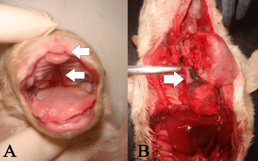
Figure 1
(A) Cleft lip and cleft palate in a canine neonate. (B) Hypoplasia of ribs in a canine neonate. A single bony protuberance (arrow) is observed in the costal arcs.
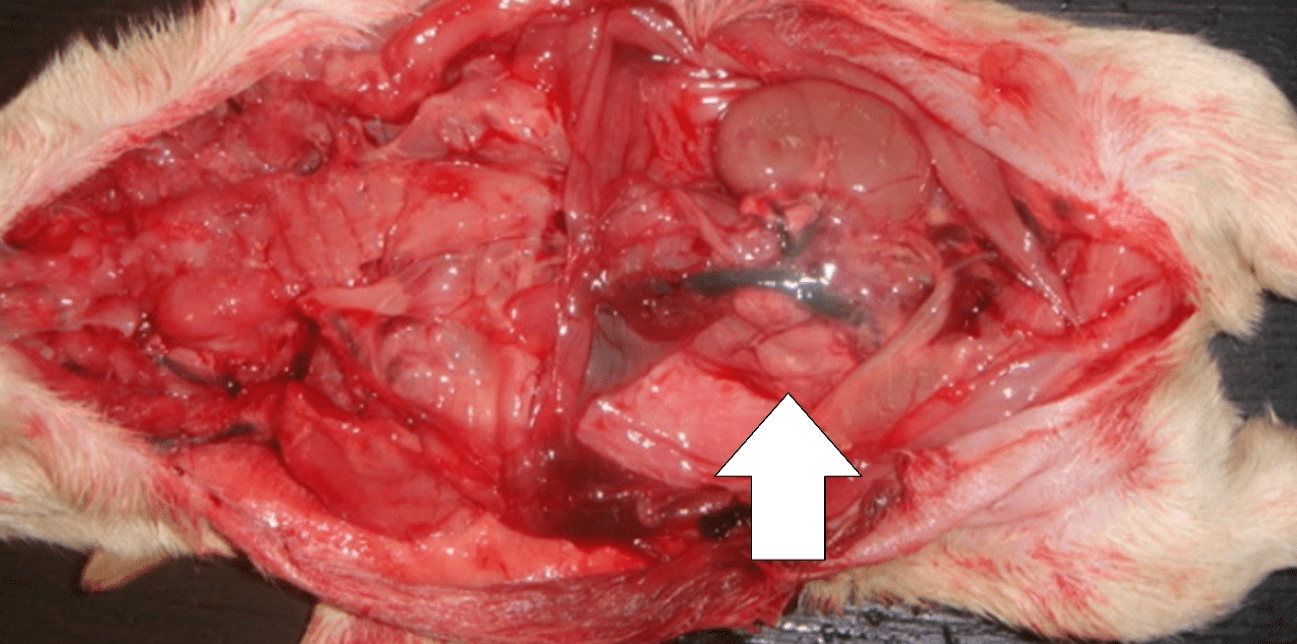
Figure 2
Right unilateral renal agenesis in a canine neonate.
Discussion
In medicine, hypoplasia of ribs is a malformation that causes respiratory distress in newborns soon after birth. It makes thoracic expansion for respiration impossible (VRIES et al., 2010). This way, the canine neonate had reduced pulmonary compliance and, thus, large part of the lungs remained collapsed. Consequently, the neonate exhibited severe dyspnoea and cyanosis, and soon thereafter apnoea, which could progress to a cardiorespiratory arrest, thus making this congenital defect incompatible with life. For this reason we chose to perform the euthanasia of the animal.
There have been reports of breed-related genetic predisposition for cleft palate, cleft lip (PETERSON; KUTZLER, 2011), and unilateral renal agenesis (HOSKINS, 2001; FUJITA et al., 2016). The canine neonate exhibited these malformations. However, it should be considered that, even of undefined breed, an animal may receive genetic characteristics from an ancestor of defined breed that may have been predisposed to these anomalies (FALCONER; MACKAY, 1996). It was not possible to assess the genealogical history of the animal. It was known that the mother was of undefined breed, but the father and/or the grandparents could have been of defined breed and transmitted the genetic characteristics of these malformations. Some genetic disorders may still skip generations (GOUGH; THOMAS, 2011). The mother of the canine newborn did not exhibit malformations, but could have carried a genetic particularity that manifested in her pup.
In medicine, hypoplasia of ribs has been reported for short rib syndrome and polydactyly, which is a rare and lethal condition of autosomal recessive inheritance. Its prevalence is related to consanguinity (DAGONEAU et al., 2009), given that endogamy increases the chances of the child to receive genes for this defect (FALCONER; MACKAY, 1996; GOUGH; THOMAS, 2011). It was not possible to determine a relationship between the father and the mother of the newborn, given that there was no information about the father and mating; however, this condition can be inherited with or without inbreeding. This genetic possibility, as the cause of the malformations in the canine neonate, is of great relevance. In the same way that it was described in the present report, hypoplasia of ribs in human neonates is also associated with other malformations, such as cleft palate, cleft lip, and urogenital defects, as well as malformations in other organs (DAGONEAU et al., 2009).
The dog was healthy according to clinical evaluation, blood examination, and biochemical tests (urea, creatinine, alanine aminotransferase, aspartate aminotransferase and alkaline phosphatase). During pregnancy, there was no maternal exposure to toxins, drugs, irradiation, infectious diseases, or traumas that could suggest a teratogenic factor involved in the malformations found.
Therefore, the cause of congenital defects in this newborn was possibly related to a hereditary genetic factor. The prevention of genetic abnormalities in dogs includes the development of genetic improvement programmes for reproduction, genetic testing of the puppies and the parents, the assessment of whether an individual is homozygous or heterozygous, and endogamy testing, which may be critical for avoiding malformations and losses in the litters (LOURENÇO, 2015; CASAL, 2016).
There are defects of unknown origin; however, the causes can be investigated based on the history of the bitches, mating, and pregnancy. Specific DNA tests are available for detecting some hereditary diseases. Genetic identification is very promising for the elimination of genetic diseases in dogs; however, there are barriers, such as limited availability of tests and their use to a large extent for unique gene disorders, which demonstrate the need for screening programmes oriented to the elimination of inherited disorders (GOUGH; THOMAS, 2011; LOURENÇO, 2015).
Conclusions
Hypoplasia of ribs is a malformation incompatible with life. It makes thoracic expansion and pulmonary compliance impossible, causing respiratory distress and neonatal mortality shortly after birth.
References
CASAL, M. L. Congenital and genetic diseases of puppies before the weaning: can we prevent them? In: INTERNATIONAL SYMPOSIUM ON CANINE AND FELINE REPRODUCTION, 8., 2016, Paris. Proceedings [...]. Paris: [s.n.], 2016. p. 46.
DAGONEAU, N.; GOULET, M.; GENEVIÈVE, D.; SZNAJER, Y.; MARTINOVIC, J.; SMITHSON, S.; HUBER, C.; BAUJAT, G.; FLORI, E.; TECCO, L.; CAVALCANTI, D.; DELEZOIDE, A. L.; SERRE, V.; LE MERRER, M.; MUNNICH, A.; CORMIER-DAIRE, V. DYNC2H1 mutations cause asphyxiating thoracic dystrophy and short rib-polydactyly syndrome, type III. American Journal of Human Genetics, Houston, v. 84, n. 5, p. 706-711, 2009.
DUONG, A.; STEINMAUS, C.; MC-HALE, C. M.; VAUGHAN, C. P.; ZHANG, L. Reproductive and developmental toxicity of formaldehyde: a systematic review. Mutation Research, Amsterdam, v. 728, n. 3, p. 118-138, 2011.
FALCONER, D. S.; MACKAY, T. F. C. Introduction to quantitative genetics. 4th ed. Harlow: Longman Group Limited, 1996. 480 p.
FUJITA, A.; TSUBOI, M.; UCHIDA, K.; NISHIMURA, R. Complex malformations of the urogenital tract in a female dog: gartner duct cyst, ipsilateral renal agenesis, and ipsilateral hydrometra. Japanese Journal of Veterinary Research, Sapporo, v. 64, n. 2, p. 147-152, 2016.
GILL, M. A. Perinatal and late neonatal mortality in the dog. 2001. Thesis (Doctor of Philosophy) -University of Sydney, New South Wales, 2001.
GOUGH, A.; THOMAS, A. Breed predispositions to disease in dog and cats. 2th ed. Hoboken: Wiley-Blackwell, 2011. 352 p.
HOSKINS, J. D. Veterinary pediatrics: dogs and cats from birth to six months. 3th ed. Philadelphia: Saunders, 2001. 594 p.
LOURENÇO, M. L. G. Cuidados com neonatos e filhotes. In: JERICÓ, M. M.; KOGIKA, M. M.; ANDRADE NETO, J. P. (Ed.). Tratado de medicina interna de cães e gatos. Rio de Janeiro: Roca, 2015. p. 363-475.
PETERSON, M. E.; KUTZLER, M. A. Small animal pediatrics. Saint Louis: Elsevier, 2011. 448 p.
SCHOENEBECK, J. J.; OSTRANDER, E. A. Insights into morphology and disease from the dog genome Project. Annual Review of Cell Developmental Biology, Palo Alto, v. 30, n. 1, p. 535-560, 2014.
VRIES, J.; YNTEMA, J. L.; VAN DIE, C. E.; CRAMA, N.; CORNELISSEN, E. A. M.; HAMEL, B. C. J. Jeune syndrome: description of 13 cases and a proposal for follow-up protocol. European Journal of Pediatrics, Berlim, v. 169, n. 1, p. 77-88, 2010.
Author notes
maria-lucia.lourenco@unesp.br