ARTICLES
Dynamics of hematological parameters in female lambs during the first four months of life
Dinâmica dos parâmetros hematológicos em cordeiras durante os primeiros quatro meses de vida
Dynamics of hematological parameters in female lambs during the first four months of life
Semina: Ciências Agrárias, vol. 39, no. 6, pp. 2465-2476, 2018
Universidade Estadual de Londrina
Received: 18 December 2017
Accepted: 12 April 2018
Abstract: Important physiological changes affect the blood profile of ruminants during the growth phase, but few studies approach the factors involved in these dynamics in lambs. The aim of this study was to characterize the dynamics of hematological parameters, of total plasma protein (TPP), and of fibrinogen in healthy female lambs during the first four months of life. Blood samples of 35 female lambs were collected at 30, 60, 90, and 120 days old to perform the complete blood count (CBC). The erythrocyte and leukocyte parameters, TPP, and fibrinogen were determined. The means for total red blood cell (RBC) counts at 60 and 120 days differed (P < 0.05) from the initial mean, showing a peak of 13.6 x 10⁶ cells µL-1 at 60 days old. The mean values for packed cell volume (PCV) and hemoglobin (Hgb) concentration increased (P < 0.05) until 90 days and decreased at 120 days (36.6% to 33.7% and 11.4 g dL-1 to 10.6 g dL-1 between 90 and 120 days, respectively). The means for mean corpuscular volume (MCV) and for mean corpuscular hemoglobin concentration (MCHC) increased (P < 0.05) between 30 and 120 days (27.5 µm3 to 29.7 µm3 and 26.6% to 31.4%, respectively). The total white blood cell (WBC) count increased (P < 0.05) and reached a peak at 90 days (9,314 cells µL-1). The peaks for segmented neutrophils (5,141 cells µL-1) and lymphocyte counts (4,236 cells µL-1) occurred at 60 and 90 days, respectively. The means for neutrophil/lymphocyte ratio were similar (P > 0.05) between the ages (mean of 1.8) but higher than the reference value for adult sheep (0.5). The initial mean for eosinophil counts (2 cells µL-1) was lower (P < 0.05) than all subsequent ones, and the monocyte count showed the lowest level (P < 0.05) at 120 days (232 cells µL-1). The mean for TPP at 60 days (6.4 g dL-1) was higher (P < 0.05) than the other ages. Except for band neutrophil and basophil counts, and fibrinogen concentration, the hematological parameters and the TPP of female lambs are influenced by age until four months of life and differ from the reference intervals established for adult sheep. Therefore, the interpretation of CBCs performed in female lambs should be made on the basis of age group-specific reference intervals.
Key words: Age, Erythrogram, Fibrinogen, Leukogram, Neutrophil/Lymphocyte Ratio, Sheep.
Resumo: Importantes alterações fisiológicas influenciam o perfil sanguíneo dos ruminantes durante a fase de crescimento, mas poucos estudos abordam os fatores envolvidos nessa dinâmica em cordeiros. O objetivo deste estudo foi caracterizar a dinâmica dos parâmetros hematológicos, da proteína plasmática total (PPT) e do fibrinogênio em cordeiras saudáveis durante os primeiros quatro meses de vida. Foram coletadas amostras de sangue de 35 cordeiras aos 30, 60, 90 e 120 dias de idade para a realização do hemograma. Os parâmetros eritrocitários e leucocitários, a PPT e o fibrinogênio foram determinados. As médias da contagem total de eritrócitos aos 60 e 120 dias diferiram (P < 0,05) da média inicial, apresentando pico de 13,6 x 10⁶ células µL-1 aos 60 dias de idade. Os valores médios do volume globular e da concentração de hemoglobina aumentaram (P < 0,05) até os 90 dias e diminuíram aos 120 dias (36,6% para 33,7% e 11,4 g dL-1 para 10,6 g dL-1 entre 90 e 120 dias, respectivamente). As médias de volume globular médio (VGM) e de concentração de hemoglobina globular média (CHGM) aumentaram (P < 0,05) entre 30 e 120 dias (27,5 µm3 para 29,7 µm3 e 26,6% para 31,4%, respectivamente). A contagem total de leucócitos aumentou (P < 0,05) e atingiu o pico aos 90 dias (9.314 células µL-1). Os picos para contagem de neutrófilos segmentados (5.141 células µL-1) e linfócitos (4.236 células µL-1) ocorreram aos 60 e 90 dias, respectivamente. As médias para a razão neutrófilo: linfócito foram semelhantes (P > 0,05) entre as idades (média de 1,8), porém superiores ao valor de referência para ovinos adultos (0,5). A média inicial do número de eosinófilos (2 células µL-1) foi menor (P < 0,05) que todas as posteriores, e o número de monócitos apresentou o nível mais baixo (P < 0,05) aos 120 dias (232 células µL-1). A média de PPT aos 60 dias (6,4 g dL-1) foi superior (P < 0,05) às demais idades. Com exceção da contagem de neutrófilos bastonetes, basófilos e da concentração de fibrinogênio, os parâmetros hematológicos e a PPT de cordeiras são influenciados pela idade até os quatro meses de vida, e diferem dos intervalos de referência estabelecidos para ovinos adultos. Portanto, a interpretação de hemogramas realizados em cordeiras deve ser feita com base em intervalos de referência específicos para essa faixa etária.
Palavras-chave: Eritrograma, Fibrinogênio, Idade, Leucograma, Ovino, Razão Neutrófilo: Linfócito.
Introduction
Hematological tests are essential to assist in the clinical diagnosis of diseases in ruminants and can be considered an important complement to physical examination (JONES; ALLISON, 2007). The proper interpretation of the complete blood count (CBC), combined with the animal’s medical history, assist in the differential diagnosis of various diseases and, in many cases, provides a more accurate prognosis and the choice of the best treatment strategy (POLIZOPOULOU, 2010). To turn this feasible, the prior knowledge about the reference values of the blood parameters in healthy animals is essential as well as on factors that act on its dynamics, even in the absence of diseases, which allows to discern whether or not an animal has physiological and/or pathological changes (BIRGEL JÚNIOR et al., 2001; AYRES et al., 2009).
In the late 1970s, Fan and Schons (1978) established hematological values for 100 healthy adult sheep raised on pasture in the municipality of Santa Maria, Rio Grande do Sul, Brazil. Since then, only in the last 15 years, there has been interest from researchers in the search for the establishment of reference intervals for different age groups and stages of the reproductive cycle of the Brazilian sheep flock (FERREIRA et al., 2003; BRITO et al., 2006; GAMA et al., 2007; BATISTA et al., 2009; SANTANA et al., 2009; DAVID et al., 2012; BEZERRA et al., 2013; MADUREIRA et al., 2013; LIMA et al., 2015; MENEGHINI et al., 2016). Nevertheless, specific and detailed studies on the dynamics of the hematological parameters during the first months of life are still scarce. For this reason, the reference intervals often used in the evaluation of lambs are still based on values determined for adult animals.
As observed in beef calves (BIRGEL JÚNIOR et al., 2001), dairy calves (GALINDO et al., 2009), and goat kids (AYRES et al., 2009), important physiological changes affect the blood profile of the ruminants during the growth phase because of hematopoietic and metabolic processes that occur from birth to adulthood. Therefore, a deeper understanding about the factors involved in this dynamic in lambs is necessary.
There is also a shortage of studies on the neutrophil/lymphocyte ratio in sheep. Besides being a predictive method for diseases and infections, this evaluation correlates the leukocyte profile with the levels of glucocorticoid hormones, allowing to verify the stress response and animal welfare (DAVIS et al., 2008).
In this context, the purpose of this study was to characterize the dynamics of hematological parameters, of total plasma protein (TPP), and of fibrinogen in healthy female lambs according to age development during the first four months of life.
Material and Methods
The experimental protocol (016/2011 of August 04, 2011) was approved by the Ethics Committee on Animal Use of the Agricultural Sciences Campus of the Federal University of Paraná (Universidade Federal do Paraná - UFPR, Brazil.
The study was carried out at the Sheep and Goat Production and Research Center (Laboratório de Produção e Pesquisa de Ovinos e Caprinos - LAPOC) of UFPR, located in the municipality of Pinhais, Paraná State, Southern Brazil (25º38’ S, 49º14’ W, and at 953 m altitude; in a Cfb type climate for the Köppen-Geiger classification, corresponding to the temperate maritime humid climate with temperate summer). Thirty-five ½ White Dorper x ½ Suffolk female lambs, monitored from birth to 120 days old, were used. The evaluation period extended from October to March (Spring/Summer).
As prophylactic measures, there was certified that the colostration was adequate, and the postpartum umbilical disinfection was performed as well as disinfection of the premises; vaccinations against clostridiosis (Sintoxan® Polivalente T, Merial) and ecthyma contagiosum (Ectima Vac®, CEVA) were made at 41 days old, on average; and weighings, body condition scoring (BCS) according to Russel et al. (1969), and parasitological monitoring (by observation of clinical signs such as diarrhea and the application of the Famacha method according to Molento et al. (2004)) were performed at 14-day intervals. Because of the initial small size of the animals, there was difficulty to obtain the required amount of feces for the parasitological exams and, therefore, the count of eggs per gram of feces (EPG) was not made. No complication was observed after these interventions, and only animals with BCS ≥ 2, Famacha ≤ 3, and no lesions or apparent pathological signs were considered healthy and included in the analyses.
The female lambs remained housed with their mothers in collective pens from birth to weaning, which occurred between 70 and 80 days old. From 14 days old, they had access to food in creep-feeding composed of corn silage and concentrate, provided as total mixed ration (TMR), twice a day (at 08h00 and 16h00). The protein-energetic concentrate had 214 g kg-1 of crude protein (CP) and 773.2 g kg-1 of total digestible nutrients (TDN) (consisting of 574 g kg-1 of ground corn, 134 g kg-1 of soybean hulls, 261 g kg-1 of soybean meal, 13 g kg-1 of limestone, and 18 g kg-1 of mineral supplement), and the silage had 71.8 g kg-1 of CP and 655.7 g kg-1 of TDN, based on the dry matter (DM). The diet of the ewes was composed of the same silage provided to the female lambs plus concentrate with 189.7 g kg-1 of CP and 769.2 g kg-1 of TDN (consisting of 504 g kg-1 of ground corn, 287 g kg-1 of soybean meal, 160 g kg-1 of wheat bran, 24 g kg-1 of limestone, and 25 g kg-1 of mineral supplement), adjusted for the category of lactating ewes according to National Research Council - NRC (2007). At the age of 61 days, the adjustments for feeding of the female lambs were started (Table 1), allowing leftovers of up to 20% of the fresh matter (FM) offered to not limit the feed intake.
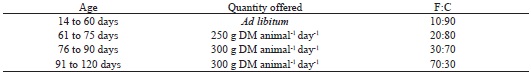
DM: dry matter; F:C: forage:concentrate ratio.
After weaning, the female lambs remained housed in collective pens until reach 90 days old. From this age, they were allocated to limpograss pastures (Hemarthria altissima; 128.8 g kg-1 of CP and 757.5 g kg-1 of TDN on a DM basis) during the daytime (from 08h00), being gathered in the fold in the evening (after 16h00) where they received supplementation in the trough (of the same composition of TMR provided in the creep-feeding during pre-weaning) and remained overnight. The diets were formulated to meet the nutritional requirements recommended for growing female lambs, with maturity of 30% at four months old, according to NRC (2007).
The CBCs were performed at 30, 60, 90, and 120 days of age. Blood samples (2 mL) were collected by jugular venipuncture in vacuum tubes containing anticoagulant (K3-EDTA 10%), after a 10-hour fast (without water deprivation). The samples were homogenized and cooled (4 °C) until analysis, which did not exceed the maximum period of 24 hours after collection. The tests were performed in the Veterinary Clinical Pathology Laboratory of UFPR.
In the erythrograms, the total red blood cell (RBC) counts and the hemoglobin (Hgb) concentrations were determined using a hematology analyzer (BC-2800 Vet, Myndray®). The packed cell volume (PCV) values were determined by microhematocrit technique (FARRAND, 1976) using capillary tubes and centrifugation at 11,000 RPM for 8 minutes. The hematimetric indices, mean corpuscular volume (MCV) and mean corpuscular hemoglobin concentration (MCHC), were calculated according to the methodology described by Wintrobe (1990).
The total white blood cell (WBC) counts were performed in duplicate in a Neubauer improved chamber. The blood samples were diluted in acetic acid 4% in a 1:20 ratio. The leukocyte differential count was performed by a trained technical team, analyzing 100 leukocytes on slides with blood smears prepared within two hours of blood collection and stained by the Quick Panoptic method (Instant Prov, Newprov®). The neutrophil/lymphocyte ratio was also calculated.
Furthermore, the TPP concentration was determined by refractometry, and the plasma fibrinogen was determined by the heat precipitation method (KANEKO; SMITH, 1967).
As the data were obtained from the same individuals at multiple moments over time (30, 60, 90, and 120 days), the linear mixed model was used to determine the mean values and standard errors of the variables evaluated. The time was established as a fixed effect, and each individual observed at each moment over time was considered as a random variable according to the model: Ŷijkl = µ + αi + βj + Ɣk + εijkl, where: Ŷijkl = value of the hematological parameter for a certain age; µ = intercept or mean of age of the female lambs at 30 days; αi = effect of age at 60 days; βj = effect of age at 90 days; Ɣk = effect of age at 120 days; and εijkl = error associated with the value of the observed parameter. The means for each age were compared over the experimental period considering the individual variation of the female lambs at a 5% significance level.
To determine the interval of the hematological parameters of the female lambs between 30 and 120 days old, the lower and upper limits of the 95% confidence interval, the median, the mean, and the standard deviation (M ± SD) were established through descriptive analysis.
Statistical analysis was performed using the computing environment R Project for Statistical Computing, version 2.10.1 (R DEVELOPMENT CORE TEAM, 2009).
Results and Discussion
The female lambs presented growth within the recommended by NRC (2007) for this category. The mean and standard error (M ± SE) for body weight at 30 and 120 days old, and for average daily gain over the period were 12.4 ± 0.5 kg, 30.5 ± 1.7 kg, and 240 ± 24 g day-1, respectively. Also, the means for Famacha grade and BCS were 2 ± 0.1 and 3 ± 0.1, respectively, indicating that the female lambs presented adequate sanitary and nutritional status, which resulted in good performance during the experiment.
There was an effect of age on all erythrocyte parameters (Table 2). The means for total RBC counts at 60 and 120 days differed (P < 0.05) from the initial mean. The peak of 13.6 x 10⁶ RBCs µL-1 occurred at 60 days old, gradually decreasing during the experimental period.
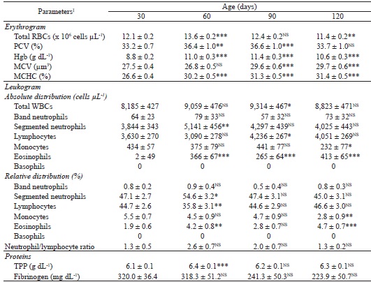
I RBC: red blood cell; PCV: packed cell volume; Hgb: hemoglobin; MCV: mean corpuscular volume; MCHC: mean corpuscular hemoglobin concentration; WBC: white blood cell; TPP: total plasma protein.* significant difference (P < 0.05) in relation to the mean at 30 days of age;** significant difference (P < 0.01) in relation to the mean at 30 days of age;*** significant difference (P < 0.001) in relation to the mean at 30 days of age; NS: non-significant difference in relation to the mean at 30 days of age (P > 0.05).
There was an increase (P < 0.05) in PCV until 90 days old, where the highest value for this parameter (36.6%) was observed, followed by a fall until 120 days old (33.7%) to a value similar (P > 0.05) to the initial (33.2%; Table 2). In parallel, the means for Hgb concentrations increased (P < 0.05) from 8.8 g dL-1 to 11.4 g dL-1 between 30 and 90 days old, decreasing (P < 0.05) to 10.6 g dL-1 at 120 days old.
These variations converge to a dynamic profile similar to those described by Ullrey et al. (1965a), Gama et al. (2007) and Bórnez et al. (2009). Ullrey et al. (1965a) when assessing Hampshire, Shropshire, and Suffolk lambs from birth to one year old found that the total RBC counts increased from 11.39 x 10⁶ µL-1 at 30 days to the maximum value of 12.95 x 10⁶ µL-1 at 90 days, followed by a gradual decrease until eight months old. These authors also observed an increase in PCV and Hgb concentration between 30 and 90 days old, in which its maximum values were reached, followed by a decrease until 150 days. The increase in the values of these parameters until 90 days old resulted from increased hematopoietic activity of the red bone marrow during the first weeks of life (JAIN, 1993).
In contrast, the decrease in the total RBC count, PCV, and Hgb concentration verified at 120 days reflected the reduction in the demand for RBCs with advancing age. In this process, the red bone marrow is invaded partially by adipose cells, which is now called yellow bone marrow and becomes inactive. The adipose tissue becomes predominant while the hematopoietic cells decrease, causing a reduction of the total erythrocyte mass (FERREIRA et al., 2003; GALINDO et al., 2009; LIMA et al., 2015). This decrease is also related to the approximation of age to puberty of the female lambs, because as there is an increase in the release of estrogens by the organism, the synthesis of erythropoietin is inhibited, which is the main humoral factor linked to the stimulation of erythropoiesis (PIERAGOSTINI et al., 2000; FERREIRA et al., 2003).
Another factor that should be considered is nutritional management, since the total RBC count, PCV, and Hgb concentration increased with the intensification of the intake of solid foods in the diet. On the other hand, the decrease in the values of these parameters after weaning suggest that the restriction of milk intake, the breakdown of maternal-filial bond, and the adaptation and learning period for grazing food search led to a temporary decrease in nutrient intake and, consequently, to lower erythropoietic activity, similar to that reported in goat kids by Shah et al. (2010).
As for the hematimetric indices, the MCV did not differ (P > 0.05) between 30 and 60 days (mean of 27.15 µm3) but increased (P < 0.05) and became stable at 90 and 120 days old (mean of 29.65 µm3; Table 2). Meanwhile, the MCHC values at 60, 90, and 120 days (ranging from 30.2% to 31.4%) were higher (P < 0.05) than the value observed at 30 days old (26.6%).
Opposing the present study, Ullrey et al. (1965a) and Gama et al. (2007) reported decreases in MCV values with advancing age in sheep. This is expected, since RBCs of fetal origin are replaced by adult cells of smaller diameters. Lima et al. (2015) found that lambs from three to six months of age may present lower values for MCV associated with higher RBC counts compared to older sheep, because the presence of smaller cells would compensate for the greater amount released into the bloodstream.
In this study, the lowest MCV values observed at 30 and 60 days old were, probably, representative of the last phase of a period of reduction of this parameter, which started in the first weeks of the neonatal period and after 60 days gave way to a new period of increase these values. These fluctuations occur in the first year of life until they stabilize between the reference values established for adults, as verified by Egbe-Nwiyi et al. (2000). The MCHC variation was directly related to the dynamics of Hgb concentration regarding age of the female lambs. These parameters were probably influenced by the increasing iron input promoted by increased intake of solid foods over the experimental period, mainly after weaning. In fact, young animals receiving only milk develop iron deficiency and present with a decrease in MCV and MCHC (WEISS, 2010).
Regarding the leukocyte profiles, it was verified that the total WBC count increased (P < 0.05) from 8,185 WBCs µL-1 to 9,314 WBCs µL-1 between 30 and 90 days, decreasing (P < 0.05) to 8,823 WBCs µL-1 at 120 days old (Table 2). This result coincided with the dynamics observed in lambs by Ullrey et al. (1965b), who observed an increase from 7,892 WBCs µL-1 to the maximum value of 9,525 WBCs µL-1 with a slight decrease to 9,097 WBCs µL-1 at 30, 90, and 150 days old, respectively. In both cases, the variation in the total WBC count with age reflects the adaptability process of the hematopoietic system to extrauterine life, which, with the advancement of age, brings the leukocyte values of young animals closer to the ideal levels for adults (JAIN, 1993). In fact, the immune competence against new challenges depends on the age and is developed slowly after birth, as the animals come across many antigens for the first time and the organs associated with the immune system become fully functional (BRUN-HANSEN et al., 2006; BÓRNEZ et al., 2009; DAVID et al., 2012).
The peaks for segmented neutrophils (5,141 cells µL-1, 54.6%) and absolute lymphocyte counts (4,236 cells µL-1) occurred at 60 and 90 days, respectively (Table 2). In the other ages, these parameters have not changed (P > 0.05) and presented mean values of 4,055 cells µL-1 and 3,590 cells µL-1 for segmented neutrophils and lymphocytes, respectively. However, a decrease (P < 0.05) was observed in the relative lymphocyte count at 60 days old (Table 2).
There was an increasing trend in lymphocyte counts between 60 and 120 days old, which, as in calves, is attributed to the physiological adaptation and maturation of the organs of the immune system, mainly those responsible for lymphocyte maturation during the neonatal phase (BIONDO et al., 1998). In general, ruminants have a leukocyte profile predominantly neutrophilic during the first months of life and, as they get older, there is increased demand and increased production of lymphocytes, which then become the cells expressed in greater proportionality (JONES; ALLISON, 2007). For this reason, it is common to observe neutrophil/lymphocyte ratios of 1:2 in adult ruminants (POLIZOPOULOU, 2010).
Although the means for neutrophil/lymphocyte ratio of the female lambs did not differ (P > 0.05) among the evaluated ages (mean of 1.8; Table 2), these were superior to those described by other researchers (FAN; SCHONS, 1978; LEPHERD et al., 2009). However, the results of the present study agree with those found by Lima et al. (2015), who when evaluating lambs aged three to 24 months also recorded values greater than 1 for the neutrophil/lymphocyte ratio in all age groups studied, which is a consequence of maintaining a high neutrophil count.
As the neutrophil/lymphocyte ratio is applied as an indicator of stress and morbidity (CAPPEL et al., 1998), the high values presented in this study may have been caused by the stress of the animals due to handling for restraint, weighings, vaccinations, changes in feed, weaning, and movement of people, among other factors. Such procedures may have been perceived by the female lambs as risks to their physical integrity, survival, and welfare, resulting in a discrepancy of the values in comparison to the values of adult animals, which are already adapted to the management system in the farm. Many studies show that several procedures performed in the usual management of production animals alter the welfare indices of the younger ones, interfering with their hematological, hormonal, and biochemical parameters (NAPOLITANO et al., 1995; BÓRNEZ et al., 2009).
The eosinophil counts were also influenced (P < 0.05) by age, in which the values found between 60 and 120 days in the absolute count (ranging from 265 cells µL-1 to 413 cells µL-1) and at 60 and 120 days in the relative count (mean of 4.45%) were higher than the values recorded at 30 days old (2 cells L-1 and 1.9%; Table 2). Ullrey et al. (1965b) also verified low values for this leukocyte type, which were similar to the values found for lambs free of parasites. Eosinophils act in defense against parasitic infections by helminths, in allergic diseases, and for the modulation of inflammatory responses in hypersensitivity reactions (YOUNG; MEADOWS, 2010). Due to historical problems of parasitic resistance in sheep farming, there is a strong relationship between the increase in the eosinophil counts and the advancement of age during the growing phase. Thus, as they develop, the female lambs become more susceptible and exposed to parasitic action, which leads to an increase in their eosinophil counts (BORJESSON et al., 2000; LEPHERD et al., 2009; LIMA et al., 2015).
The monocyte counts kept within the reference interval for adult sheep throughout the experiment (mean of 370 cells µL-1 and 4.38%; Table 2), remaining constant until 90 days and decreasing (P < 0.05) at 120 days old (232 cells µL-1 and 2.8%; Table 2). It is suggested that the reduction of monocytes in lambs of different racial groups is correlated with decreased adaptability and immune defense mechanisms because of greater sensitivity to pathogens (BINEV et al., 2006). However, it is normal that in adequate sanitary conditions the number of monocytes released into the bloodstream is at baseline levels (BATISTA et al., 2009; DAVID et al., 2012; MADUREIRA et al., 2013).
There was no effect of age (P > 0.05) on band neutrophil and basophil counts, which presented very low values (mean of 68 cells µL-1 and 0.75%) and null, respectively, during the experimental period (Table 2). The increase of these classes is expected in cases of pathological processes (JONES; ALLISON, 2007). However, healthy ruminants show low counts of these cells at all ages (BATISTA et al., 2009; MADUREIRA et al., 2013), which confirms the good health condition of the animals used in this study.
The TPP concentration is sensitive to changes in diet, but its increase (P < 0.05) at 60 days old (6.4 g dL-1; Table 2) probably occurred in response to the increase of immunoglobulins by antigenic stimulation after the vaccination of the female lambs, as described by Jain (1993).
Fibrinogen is an acute-phase protein produced by the liver that represents around 5% of TPP and acts on the mechanism of homeostasis, providing a substrate for fibrin formation and assisting in tissue repair (MURATA et al., 2004; BASTOS et al., 2016). The determination of fibrinogen concentration is complementary to the ruminant’s leukogram and is often a better indicator of inflammation than the alterations in the leukocyte profile itself. The increase in fibrinogen indicates acute inflammatory processes, bacterial infections, surgical trauma, tissue injuries, and cases of dehydration, while its decrease occurs in liver diseases, in cases of disseminated intravascular coagulation, and to prove the success of therapeutic treatments in ruminants (MURATA et al., 2004; JONES; ALISSON, 2007; SANTOS et al., 2014). In this study, the fibrinogen concentration was not influenced by age (P > 0.05) and kept constant (mean of 275.9 mg dL-1; Table 2), reinforcing the good health condition of the female lambs.
The Table 3 presents the results of descriptive statistics of hematological parameters, of TPP concentration, and of fibrinogen of the female lambs that were assessed in this study, and its comparison with the reference interval for adult sheep established by Kramer (2000). Although it refers to a small and homogeneous sample population, the results of healthy animals in the age group between 30 and 120 days can aid in the interpretation of diagnostic tests of other animals in this phase.
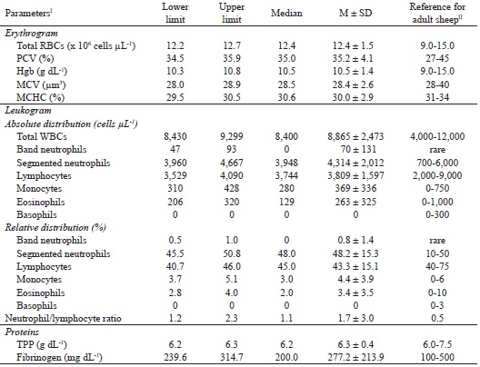
I RBC: red blood cell; PCV: packed cell volume; Hgb: hemoglobin; MCV: mean corpuscular volume; MCHC: mean corpuscular hemoglobin concentration; WBC: white blood cell; TPP: total plasma protein.II Source:Kramer (2000).
In general, the erythrocyte parameters maintained values close to the lower limit of the reference interval for adult sheep. The mean and the limits for MCV were very close to the lower limit, while the mean and the limits for MCHC were below the lower limit of the reference interval for adult sheep (Table 3).
In the leukograms, the mean for segmented neutrophils added to its standard deviation (6,326 cells µL-1, 63.5%; Table 3) exceeds the upper limit of the reference interval for adult animals. The lymphocyte values, although within the reference interval for adults, maintained close to its lower limit, so that its mean in relative distribution subtracted from its standard deviation (28.2%) was below the mean of the reference interval (Table 3). The mean for monocytes added to its standard deviation in relative distribution (8.3%) requires attention, because although it was higher than the reference values for adults, it remained within the reference limits in its absolute distribution (Table 3), which has greater accuracy for interpretation of exams. Eosinophil values remained within the reference interval for the species but close to its lower limit (Table 3). The mean and the limits for the neutrophil/lymphocyte ratio were higher than those reported for adults, with the mean being about 3.4 times higher than the reference mean (1.7 vs. 0.5; Table 3).
The TPP values remained close to the lower limit of the reference, so that subtracting the standard deviation from the mean of the female lambs, the concentration became lower than the reference interval for adults (5.9 g dL-1; Table 3). The fibrinogen mean subtracted from its standard deviation resulted in 63.3 mg dL-1 (Table 3), so female lambs between 30 and 120 days may present values lower than 100 mg dL-1, which would not be considered normal in adult sheep.
The results of this study confirm that the hematological values of younger animals often differ from the reference intervals for adults. As a result, it is recommended that more comprehensive studies be carried out with larger number of animals and in different geographic regions towards to establishment of reference intervals of blood parameters for lambs and adult sheep raised in Brazil.
Conclusions
Except for band neutrophil and basophil counts, and fibrinogen concentration, the hematological parameters and total plasma protein concentration of healthy female lambs are influenced by age until four months of life and differ from the reference intervals established for adult sheep. The neutrophil/lymphocyte ratio does not change during the four months but differ from that established for adulthood. Therefore, the interpretation of complete blood counts performed in female lambs should be made on the basis of age group-specific reference intervals.
Acknowledgements
Alda Lúcia Gomes Monteiro is member of the MARCARNE Network, funded by CYTED (ref. 116RT0503). The authors thank all the students and staff of the LAPOC-UFPR and of the Veterinary Clinical Pathology Laboratory of UFPR for their support.
References
AYRES, M. C. C.; DOREA, R. D.; BIRGEL JÚNIOR, E. H.; VIANA, R. B.; LARA, M. C. C. S. H.; BITTENCOURT, T. C. B. S. C.; BIRGEL, E. H. Dinâmica do leucograma de caprinos jovens, do nascimento até seis meses de idade: influência do fator racial. Ciência Animal Brasileira, Goiânia, v. 10, p. 261-265, 2009. Suplemento 1.
BASTOS, R. N.; MARQUES, T. L. P.; RAMOS, M. T.; MELLO COSTA, M. F. de. Avaliação das concentrações de fibrinogênio plasmático em equinos da raça Mangalarga Marchador: efeito do exercício, do gênero e da idade. Revista de Saúde, Vasouras, v. 7, n. 2, p. 12-15, jul./dez. 2016.
BATISTA, M. C. S.; CASTRO, R. S.; REGO, E. W.; CARVALHO, F. A. A.; SILVA, S. M. M. S.; CARVALHO, C. C. D.; RIET-CORREA, F. Hemograma, proteinograma, ionograma e dosagens bioquímicas e enzimáticas de ovinos acometidos por conidiobolomicose no Nordeste do Brasil. Pesquisa Veterinária Brasileira, Seropédica, v. 29, n. 1, p. 17-24, jan. 2009.
BEZERRA, L. R.; TORREÃO, J. N. C.; MARQUES, C. A. T.; MACHADO, L. P.; ARAÚJO, M. J.; VEIGA, A. M. S. Influência da suplementação concentrada e da categoria animal no hemograma de ovinos da raça Morada Nova. Arquivo Brasileiro de Medicina Veterinária e Zootecnia, Belo Horizonte, v. 65, n. 6, p. 1738-1744, 2013.
BINEV, R.; SLAVOVA, P.; LAVEVA, S. Effects of fasting on blood cells from lambs of various breeds. Trakia Journal of Sciences, Stara Zagora, v. 4, n. 3, p. 37-43, 2006.
BIONDO, A. W.; LOPES, S. T. A.; KOHAYAGAWA, A.; TAKAHIRA, R. K.; ALENCAR, N. X. Hemograma de bovinos (Bos indicus) sadios da raça Nelore no primeiro mês de vida, criados no Estado de São Paulo. Ciência Rural, Santa Maria, v. 28, n. 2, p. 251-256, 1998.
BIRGEL JÚNIOR, E. H. ; D’ANGELINO, J. L.; BENESI, F. J.; BIRGEL, E. H. Valores de referência do eritrograma de bovinos da raça Jersey criados no Estado de São Paulo. Arquivo Brasileiro de Medicina Veterinária e Zootecnia, Belo Horizonte, v. 53, n. 2, p. 1-9, 2001.
BORJESSON, D. L.; CHRISTOPHER, M. M.; BOYCE, W. M. Biochemical and hematologic reference intervals for free-ranging desert bighorn sheep. Journal of Wildlife Diseases, Lawrence, v. 36, n. 2, p. 294-300, abr. 2000.
BÓRNEZ, R.; LINARES, M. B.; VERGARA, H. Haematological, hormonal and biochemical blood parameters in lamb: effect of age and blood sampling time. Livestock Science, Amsterdam, v. 121, n. 2-3, p. 200-206, abr. 2009.
BRITO, M. A.; GONZÁLEZ, F. D.; RIBEIRO, L. A.; CAMPOS, R.; LACERDA, L.; BARBOSA, P. R.; BERGMANN, G. Composição do sangue e do leite em ovinos leiteiros do Sul do Brasil: variações na gestação e na lactação. Ciência Rural, Santa Maria, v. 36, n. 3, p. 942-948, maio/jun. 2006.
BRUN-HANSEN, H. C.; KAMPEN, A. H.; LUND, A. Hematologic values in calves during the first six months of life. Veterinary Clinical Pathology, Medford, v. 35, n. 2, p. 182-187, jun. 2006.
CAPPEL, T.; BUENO, A.; CLEMENS, E. Calving difficulty and calf response to stress. Nebraska Beef Cattle Reports, Lincoln, n. 327, p. 16-19, jan. 1998. Available at: <http://digitalcommons.unl.edu/animalscinber/327>. Accessed at: 7 dez. 2017.
DAVID, C. M. G.; LUQUETTI, B. C.; COSTA, R. L. D.; BONELLO, F. L. Padrão hematológico de cordeiros da raça Santa Inês criados sob manejo semi-extensivo na região Oeste do Estado de São Paulo. Boletim de Indústria Animal, Nova Odessa, v. 69, n. 1, p. 79-84, jan./jun. 2012.
DAVIS, A. K.; MANEY, D. L.; MAERZ, J. C. The use of leukocyte profiles to measure stress in vertebrates: a review for ecologists. Functional Ecology, London, v. 22, n. 5, p. 760-772, out. 2008.
EGBE-NWIYI, T. N.; NWAOSU, S. C.; SALAMI, H. A. Haematological values of appararently healthy sheep and goats as influenced by age and sex in arid zone of Nigeria. African Journal of Biomedical Research, Grahamstown, v. 3, n. 2, p. 109-115, 2000.
FAN, L. C. R.; SCHONS, J. A. B. Valores hematológicos de ovinos adultos normais no município de Santa Maria. Revista do Centro de Ciências Rurais, Santa Maria, v. 8, n. 1, p. 1-5, 1978.
FARRAND, L. L. The microhematocrit: technique and applications. The Nurse Practitioner, Philadelphia, v. 1, n. 5, p. 19-20, maio/jun. 1976.
FERREIRA, A. F.; RÊGO, E. W.; MELO, L. E. H.; MELO, M. T.; MENDES, E. I.; GALINDO, R. C. G. ; MENEZES, É. S. B. Eritrograma de ovinos (Ovis aries, LINNAEUS, 1758) da raça Santa Inês, clinicamente sadios, criados na Mesorregião Metropolitana de Recife. Influência dos fatores sexual e etário. Ciência Veterinária nos Trópicos, Recife, v. 6, n. 2-3, p. 89-95, maio/dez. 2003.
GALINDO, R. C. G.; FERREIRA, A. F.; MENDES, E. I.; SANTOS, S. B.; ANDRADE, R. L. F. S.; BATISTA, D. M.; LIMA, S. K. D.; RÊGO, E. W. Eritrograma de bovinos da raça Holandesa criados na Mesorregião Metropolitana do Recife: influência dos fatores sexual e etário. Medicina Veterinária, Recife, v. 3, n. 3, p. 1-6, jul./set. 2009.
GAMA, S. M. S.; MATOS, J. R.; ZACHARIAS, F.; CHAVES FILHO, R. M.; GUIMARÃES, J. E.; BITTENCOURT, T. C. B. S. C.; AYRES, M. C. C. Dinâmica do eritrograma de cordeiros, resultantes do cruzamento entre animais de raças nativas criadas no Nordeste e a raça Dorper, desde o nascimento até os seis meses de idade. Revista Brasileira de Saúde e Produção Animal, Salvador, v. 8, n. 1, p. 11-23, 2007.
JAIN, N. C. Essentials of veterinary hematology. Philadelphia: Lea & Febiger, 1993. 417 p.
JONES, M. L.; ALLISON, R. W. Evaluation of the ruminant complete blood cell count. Veterinary Clinics Food Animal Practice, Amsterdam, v. 23, n. 3, p. 377-402, nov. 2007.
KANEKO, J. J.; SMITH, H. The estimation of plasma fibrinogen and its clinical significance in the dog. The California Veterinarian, Sacramento, v. 21, n. 4, p. 21-24, 1967.
KRAMER, J. W. Normal hematology of cattle, sheep, and goats. In: FELDMAN, B. F.; ZINKL, J. G.; JAIN, N. C. (Ed.). Schalm’s veterinary hematology. 5th ed. Philadelphia: Lippincott Williams and Wilkins, 2000. p. 1075-1084.
LEPHERD, M. L.; CANFIELD, P. J.; HUNT, G. B.; BOSWARD, K. L. Haematological, biochemical and selected acute phase protein reference intervals for weaned female Merino lambs. Australian Veterinary Journal, Medford, v. 87, n. 1-2, p. 5-11, jan./fev. 2009.
LIMA, M. B.; MONTEIRO, M. V. B.; JORGE, E. M.; CAMPELLO, C. C.; RODRIGUES, L. F. S.; VIANA, R. B.; MONTEIRO, F. O. B.; COSTA, C. T. C. Intervalos de referência sanguíneos e a influência da idade e sexo sobre parâmetros hematológicos e bioquímicos de ovinos da raça Santa Inês criados na Amazônia Oriental. Acta Amazonica, Manaus, v. 45, n. 3, p. 317-322, set. 2015.
MADUREIRA, K. M.; GOMES, V.; BARCELOS, B.; ZANI, B. H.; SHECAIRA, C. L.; BACCILI, C. C.; BENESI, F. J. Parâmetros hematológicos e bioquímicos de ovinos da raça Dorper. Semina: Ciências Agrárias, Londrina, v. 34, n. 2, p. 811-816, mar./abr. 2013.
MENEGHINI, R. C. M.; BENESI, F. J.; HENRIQUES, L. C. S.; RIZZO, H.; MEIRA JUNIOR, E. B. S.; GREGORY, L. Hemogram of healthy sheep (Ovis aries) of the Santa Ines breed raised in the region of Piedade, São Paulo State: influence of age and sex. Brazilian Journal of Veterinary Research and Animal Science, São Paulo , 53, n. 4, p. 1-7, 2016.
MOLENTO, M. B.; TASCA, C.; GALLO, A.; FERREIRA, M.; BONONI, R.; STECCA, E. Método Famacha como parâmetro clínico individual de infecção por Haemonchus contortus em pequenos ruminantes. Ciência Rural, Santa Maria, v. 34, n. 4, p. 1139-1145, 2004.
MURATA, H.; SHIMADA, N.; YOSHIOKA, M. Current research on acute phase proteins in veterinary diagnosis: an overview. The Veterinary Journal, Amsterdam, v. 168, n. 1, p. 28-40, jul. 2004.
NAPOLITANO, F.; MARINO, V.; ROSA, G. de; CAPPARELLI, R.; BORDI, A. Influence of artificial rearing on behaviour and immune response of lambs. Applied Animal Behaviour Science, Amsterdam, v. 45, n. 3-4, p. 245-253, nov. 1995.
NATIONAL RESEARCH COUNCIL - NRC. Nutrient requirements of small ruminants: sheep, goats, cervids and new world camelids. Washington: National Academy of Sciences, 2007. 347 p.
PIERAGOSTINI, E.; PETAZZI, F.; RUBINO, G.; RULLO, R.; SASANELLI, M. Switching emoglobinico, quadro ematologico e primo incontro con i parassiti endoeritrocitari enzootici in agnelli autoctoni pugliesi. Obiettivi & Documenti Veterinari, Bologna, v. 21, n. 7-8, p. 31-40, 2000.
POLIZOPOULOU, Z. S. Haematological tests in sheep health management. Small Ruminant Research, Amsterdam, v. 92, n. 1-3, p. 88-91, ago. 2010.
R DEVELOPMENT CORE TEAM - R. The R project for statistical computing [: Free Software Programming Language]: R Project. Version 2.10.1. Vienna: R Foundation for Statistical Computing, 2009. Available at: <http://www.r-project.org>. Accessed at: 16 abr. 2018.
RUSSEL, A. J. F.; DONEY, J. M.; GUNN, R. G. Subjective assessment of body fat in live sheep. Journal Agricultural Science, Toronto, v. 72, n. 3, p. 451-454, 1969.
SANTANA, A. M.; SILVA, D. G.; BERNARDES, P. A.; PIZAURO, L. J. L.; MALUTA, R. P.; AQUINO, G. V.; GARCIA, K. O.; ÁVILA, F. A.; FAGLIARI, J. J. Hemograma e perfil bioquímico sérico de ovinos em idade de abate. Ciência Animal Brasileira, Goiânia, v. 10, p. 286-289, 2009. Suplemento 1
SANTOS, F. C. C.; FEIJÓ, L. S.; KASINGER, S.; FREY JUNIOR, F.; CURCIO, B. R.; NOGUEIRA, C. E. W. Hematologic values of thoroughbred foals from birth to six months of age. Ciência Animal Brasileira, Goiânia, v. 15, n. 3, p. 307-312, jul./set. 2014.
SHAH, F.; MEENAKSHI, G.; POONIA, J. S. Studies of different haematological parameters in male Beetal goats at different physiological stages. Haryana Veterinarian, Hisar, v. 49, n. 1, p. 38-39, dez. 2010.
ULLREY, D. E.; MILLER, E. R.; LONG, C. H.; VINCENT, B. H. Sheep hematology from birth to maturity. I Erythrocyte population, size and hemoglobin concentration. Journal of Animal Science, Champaign, v. 24, n. 1, p. 135-140, 1965a.
______. Sheep hematology from birth to maturity. II Leucocyte concentration and differential distribution. Journal of Animal Science, Champaign, v. 24, n. 1, p. 141-144, 1965b.
WEISS, D. J. Iron and copper deficiencies and disorders of iron metabolism. In: WEISS, D. J.; WARDROP, K. J. (Ed.). Schalm’s veterinary hematology. 6th ed. Iowa: Blackwell Publishing Ltd., 2010. cap. 26, p. 167-171.
WINTROBE, M. M. The size and hemoglobin content of erythrocyte. Methods of determination and clinical application - 1932. Journal of Laboratory Clinical Medicine, Amsterdam, v. 115, n. 3, p. 374-387, mar. 1990.
YOUNG, K. M.; MEADOWS, R. L. Eosinophils and their disorders. In: WEISS, D. J.; WARDROP, K. J. (Ed.). Schalm’s veterinary hematology. 6th ed. Iowa: Blackwell Publishing Ltd ., 2010. cap. 43, p. 281-289.
Author notes
*Author for correspondence