Abstract: The association between Polysiphonia spp and ovigerous females of Caligus rogercresseyi is analysed. Females carrying egg sacs exhibited individuals of Polysiphonia spp externally attached to the cuticle of both dorsal cephalothorax and abdomen by a mounting disk without penetrating the tissues.
Keywords:PolysiphoniaPolysiphonia, epibiont epibiont, Caligus Caligus, Salmo Salmo.
Resumen: Se analiza la asociación de Polysiphonia spp con hembras ovigeras de Caligus rogercresseyi. Las hembras portadoras de sacos ovígeros presentaron ejemplares de Polisiphonia spp adheridos externamente a la cutícula de la zona dorsal del cefalotórax y abdomen mediante un disco de fijación, sin penetrar en los tejidos.
Palabras clave: Polysiphonia, epibionte, Caligus, Salmo.
Polysiphonia spp as epibiont of Caligusrogercresseyi (Crustacea: Copepoda), in Salmo salar farming centers
Descripción de Polysiphonia spp como epibionte de Caligus rogercresseyi (Crustacea: Copepoda), en centros de cultivo de Salmo salar
Universidad Austral de Chile
Accepted: 31 March 2016
Lack of available space is a limiting factor for species looking for a suitable hard substratum to settle on, specially in marine subtidal habitats where soft sediments prevail (Connell and Keough 1985). The colonization of animals by benthic sessile marine organisms is a survival strategy (epibiosis) which provides the epibiont with this valuable resource (AbeIló et al 1990). Consequently a number of bacteria, diatoms, sessile colonial ciliates and macroalgae have been described as epibionts (Wahl and Mark 1999).
A number of protozoans have been cited as epibionts of the copepod Lepeophtheirus salmonis (Fernandez- Leborans et al 2005) an ectoparasite of the family Caligidae. Treasurer (2002) and Jones and Beamish (2011) have described for this parasite the presence of the alga Ulva spp, some ciliated protozoans and monogeneans of the Udonella genus. In Chile, Udonella australis has been described living on the surface of the copepods C. rogercresseyi and Lepeophtheirus mugiloidis, both parasites co-exist on the skin of the rock cod Eleginops maclovinus (Marin et al 2007).
The high prevalence and intensity on C. rogercresseyi led to argue that the dispersion process of monogenean occurs through the sexual contacts of caligids; for that reason U. australis is found at different stages of development on top of the egg sacs of ovigerous females (Marin et al 2007).
After histological analyses of the relationship between C. rogercresseyi and the epibiont, it was determined that Udonella spp adheres to the membrane lining the shell of the crustacean, without penetrating these structures, remaining fixed or moving on top of the shell and preying on the host fish mucus (Carvajal et al 1998, 2001).
Some macroalgae species have been identified by Perez-Martinez et al (2001) as epibionts of the cladoceran Daphnia pulicaria, a dominant crustacean on a lake in Spain, suggesting that the relationship came when the substrate and the algae were in high aggregations. In Caligids, only the presence of Ulva spp on L. salmonis has been reported as epibiont, however no further information regarding the interaction between the two species exists (Jones and Beamish 2011).
In this study we will report the presence of a red alga attached to some specimens of the parasite C. rogercresseyi collected in a Salmo salar farming center, in Chiloé.
Because epibionts were found only in females, a total of 200 C. rogercresseyi ovigerous females were collected on June 15, 2015 from a S. salar farming center of Marine Harvest Chile in ACS 10a neighborhood, Southeast of Lemuy Island (Chiloe). Salmon cages were located between 0 and 20 meters deep; environmental variables measured at sampling time were 6.7 mg/L O2, 76.4% O2 saturation, 11.6 °C (temperature) and 32 PSU in salinity.
Females were isolated from fishes anesthetised with Benzocaine 20%, then transferred to the Central Laboratory of Marine Harvest in a 1 liter plastic container with constant aeration in a ratio of 200 individuals per liter of sea water and keeping the temperature below 12 °C. Subsequently, a number of parasites were fixed in glutaraldehyde 3% for further analysis at electron microscopy, some other live individuals were sent to the laboratory of reproductive biology at Universidad Austral de Chile (UACh) in Valdivia, for further studies.
Ten live parasites were analysed by using a stereomicroscope LEICA EZ4D with incident light and provided with a CANON Powershot A1200 HD camera, in order to obtain photographic registry. Subsequently, six specimens were preserved in a formaldehyde solution at 5%, and later treated according to optical microscopy protocols. Histological sections of 7 µm thick were stained with hematoxylin and eosin and then observed at a LEICA CME light microscope.
Four specimens were fixed in a glutaraldehyde solution at 3% in order to conduct studies at Scanning Electron Microscopy (SEM). The specimens were post-fixed in a 1% OsO4 solution for 2 h at 4 °C, and later dehydrated and dried at critical point of CO2, then mounted on a holder, coated with a gold film and observed on a scanning electron microscope LEO-420.
The observation at stereomicroscope of ovigerous females of C. rogercresseyi allowed to establish the presence of a number of clamping discs including red filaments attached to the dorsal surface of the cephalothorax and the genital segment of the parasite (figure 1a). Near to areas of fixation discs, a significant number of growing strands with segmented appearance varying in size were observed (figure 1b). A number of prostrate and erect ecorticate axes, attaching by unicellular rhizoids in connection with pericentral cells, were observed on the genital segment of c. rogercresseyi. Spermatangial branches were observed directly from each axial cell and the mature carpogonial branch was four-celled. These observations suggested presence of a representative of filamentous red algae (Rhodophyta) Ceramiales order, Polysiphonia genus.
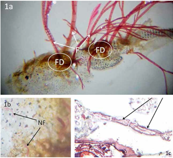
Figure 1.
a) General view of fixation discs of Polysiphonia spp in the dorsal region of an ovigerous female of Caligus rogercresseyi. FD, fixation discs; F, filaments (350X); b) new growing filaments of Polysiphonia spp on the dorsal region of Caligus rogercresseyi. NF, new filaments (1400x); c) cross section at genital level of Caligus rogercresseyi. Arrows showing cell elements of Polysiphonia spp filaments (100X).
a)Vista general de discos de fijación de Polysiphonia spp. Situados en la región dorsal de una hembra ovígera de Caligus rogercresseyi. FD, disco de fijación; F, filamentos (350X). b) nuevos filamentos de Polysiphonia spp en la región dorsal de Caligus rogercresseyi. NF, nuevos filamentos (1400x). c) sección transversal a nivel de la zona genital de Caligus rogercresseyi. Flechas muestras células que forman parte del filamentos de Polysiphonia spp (1000x).
Longitudinal and transversal histological sections at cephalothorax level showed Polysiphonia spp filaments, attached to a thick outer coating, chitinous type (figure 1c), no penetration on the parasite structures was observed.
SEM observations contributed to the identification of the algae filaments as formed by columnar cells of strands of Polysiphonia specimens. Polysiphonia spp do not penetrate the parasite, but appears attached to both the cephalothorax and genital segment through mounting discs formed by a number of rhizoids connected with pericentral cells attached to the outer coating of C. rogercresseyi (figures 1a, 2a, 2b). An abundant number of rhizoids, diatoms and also a film of bacteria in the fixation disc facilitate the germination of spores of Polysiphonia spp (figures 2b, 2c).
The literature indicates that epibionts are located at specific sites of the basibiont which meet the requirements for survival and dispersal. In algae, for example, the amount of light and the exposure to water flows are described as determining factors for development (Borowitzca et al 1990, Romagnoli et al 2007).
As previously mentioned, algae are able to grow over a number of species including crustacean (Wahl and Mark 1999) that can serve as a substrate for spore germination, growth of thallus and subsequent development of filaments, acting as epibiont.
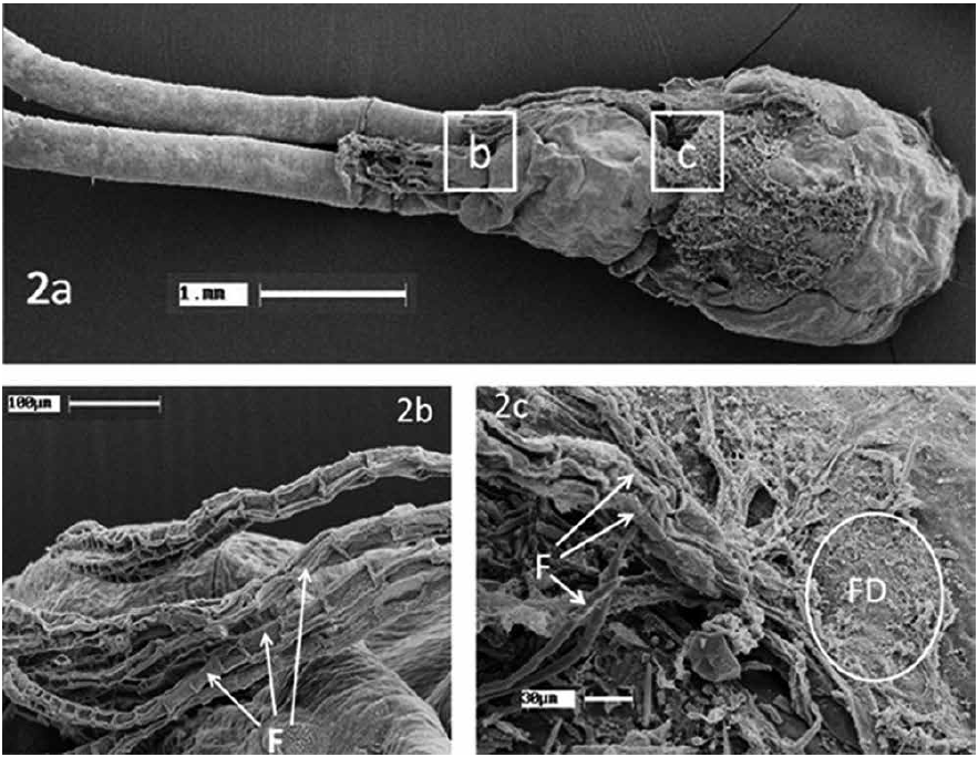
Figure 2.
a) SEM images of ovigerous female of Caligus rogercresseyi showing Polysiphonia spp filaments as epibiont. Frames b and c are magnified in figures b and c; b) filaments of Polysiphonia spp in the limit between egg capsule and genital segment of an ovigerous female of Caligus rogercresseyi. F, filaments; c) Polysiphonia spp attached to the cephalotorax of Caligus rogercresseyi. FD, fixation disc; F, filaments.
a) Imágenes a MEB de una hembra ovígera de Caligus rogercresseyi mostrando filamentos de Polysiphonia spp recuadros b y c aparecen a mayor aumento en las figuras b y c; b) filamentos de Polysiphonia spp ubicados en el límite entre la cápsula ovígera y el segmento genital de una hembra ovígera de Caligus rogercresseyi. F, filamentos; c) filamentos de Polysiphonia spp adheridos al cefalotórax de Caligus rogercresseyi. FD, discos de fijación; F, filamentos.
Because algae are primary producers in the food chain of aquatic environment, they are able to produce organic substances from inorganic substances converting the light energy into chemical energy by means of photosynthesis. Then it is possible to assume that spores of Polysiphonia spp attached to the dorsal surface of cephalotorax or in the genital region of C. rogercresseyi could be only a random event and correspond to the first substrate available for attaching, which could be associated to the presence of diatoms and also a film of bacteria facilitating the disc germination and dispersal of spores of Polysiphonia spp (Kim and Yang 2005).
Olivares et al (1998) have described the presence of filaments of Polysiphonia spp as epibiont of the limpet Fissurella latimarginata, off the coast of the north of Chile while representatives of the same algae genus have been described as fixed to the shell of some sea turtles (Baez et al 2001).
The analysis of previously reported results suggest that the spores of Polysiphonia spp use the C. rogercresseyi shell as substrate for settlement and further growth and development of the thallus, however no relationship of nutritive dependency is established because these algae are photosynthetic.
Laboratory observations suggest that attachment of Polysiphonia spp to the cephalothorax of C. rogercresseyi has no effect on the life condition or reproductive capacity of the parasite because algae do not penetrates on the parasite structures1.
Figure 1.
a) General view of fixation discs of Polysiphonia spp in the dorsal region of an ovigerous female of Caligus rogercresseyi. FD, fixation discs; F, filaments (350X); b) new growing filaments of Polysiphonia spp on the dorsal region of Caligus rogercresseyi. NF, new filaments (1400x); c) cross section at genital level of Caligus rogercresseyi. Arrows showing cell elements of Polysiphonia spp filaments (100X).
a)Vista general de discos de fijación de Polysiphonia spp. Situados en la región dorsal de una hembra ovígera de Caligus rogercresseyi. FD, disco de fijación; F, filamentos (350X). b) nuevos filamentos de Polysiphonia spp en la región dorsal de Caligus rogercresseyi. NF, nuevos filamentos (1400x). c) sección transversal a nivel de la zona genital de Caligus rogercresseyi. Flechas muestras células que forman parte del filamentos de Polysiphonia spp (1000x).
Figure 2.
a) SEM images of ovigerous female of Caligus rogercresseyi showing Polysiphonia spp filaments as epibiont. Frames b and c are magnified in figures b and c; b) filaments of Polysiphonia spp in the limit between egg capsule and genital segment of an ovigerous female of Caligus rogercresseyi. F, filaments; c) Polysiphonia spp attached to the cephalotorax of Caligus rogercresseyi. FD, fixation disc; F, filaments.
a) Imágenes a MEB de una hembra ovígera de Caligus rogercresseyi mostrando filamentos de Polysiphonia spp recuadros b y c aparecen a mayor aumento en las figuras b y c; b) filamentos de Polysiphonia spp ubicados en el límite entre la cápsula ovígera y el segmento genital de una hembra ovígera de Caligus rogercresseyi. F, filamentos; c) filamentos de Polysiphonia spp adheridos al cefalotórax de Caligus rogercresseyi. FD, discos de fijación; F, filamentos.
