
Esta obra está bajo una Licencia Creative Commons Atribución-NoComercial-SinDerivar 4.0 Internacional.
Recepción: 11 Julio 2025
Aprobación: 02 Septiembre 2025
DOI: https://doi.org/10.37135/ee.04.24.08
Abstract: A 19-year-old male patient with no significant medical history was the victim of a firearm attack, with penetrating injury to the thoracoabdominal and left popliteal region. A diagnosis of deep vein thrombosis of the popliteal vein was made, which was managed with anticoagulation. Weeks later, this resulted in a sensation of a growing mass in the popliteal fossa, accompanied by palpitations and paresthesias. He was evaluated in an outpatient clinic, where a mass was found in the popliteal region of the leg measuring approximately 4 cm, with an ultrasound diagnosis of popliteal pseudoaneurysm. Surgical treatment was indicated, which consisted of surgical resection of the pseudoaneurysm plus autologous femoropopliteal bypass with a saphenous vein, without surgical complications. Popliteal artery pseudoaneurysms are rare post-traumatic injuries that must be resolved promptly. The type of treatment depends on the size, location, and symptoms, as well as the expertise and experience of each center in open surgery.
Keywords: Trauma, Pseudoaneurysm, Popliteal Artery.
Resumen: Paciente masculino de 19 años, sin antecedentes de importancia, víctima de un ataque con arma de fuego, con lesión penetrante a nivel toracoabdominal y en la región poplítea izquierda. Se diagnosticó trombosis venosa profunda de vena poplítea manejada con anticoagulación; semanas posteriores este último resulta con sensación de masa en crecimiento a nivel de fosa poplítea, misma que se acompaña de palpitación, parestesias. Es valorado en consulta externa, donde se evidenció una masa en región poplítea de pierna de aproximadamente 4 cm, con diagnóstico ecográfico de pseudoaneurisma poplíteo, se indicó tratamiento quirúrgico que consistió en resección quirúrgica del pseudoaneurisma más bypass autólogo femoropoplíteo con vena safena sin complicaciones quirúrgicas. Los pseudoaneurismas de la arteria poplítea son lesiones postraumáticas infrecuentes que deben ser resueltas oportunamente, el tipo de tratamiento depende del tamaño, ubicación y sintomatología; así como también, la experticia de cada centro y experiencia con el manejo de cirugía abierta.
Palabras clave: trauma, pseudoaneurisma, arteria poplítea.
Post-traumatic Popliteal Pseudoaneurysm: Clinical Case Report
Pseudoaneurisma Poplíteo Postraumático: reporte de caso clínico
Authors:
María José Bahamonde Gaibor 1https://orcid.org/0009-0007-0266-2656
Diego Armando Mora Tenesaca 1https://orcid.org/0009-0005-0017-8364
Maria Belen Baño Jimenez 1https://orcid.org/0000-0002-2249-3315
Fernando Horacio Pérez Guerrero 2https://orcid.org/0000-0003-4164-1624
Pablo Arturo Suárez Hospital 1
Eugenio Espejo Hospital 2
Correspondence: María José Bahamonde G; Vascular Surgery Service; Pablo Arturo Suárez Hospital, VGC3 + W2M, Quito, 170103, Ecuador; email: majosbg@hotmail.com, telephone: 0984669054.
ABSTRACT
A 19-year-old male patient with no significant medical history was the victim of a firearm attack, with penetrating injury to the thoracoabdominal and left popliteal region. A diagnosis of deep vein thrombosis of the popliteal vein was made, which was managed with anticoagulation. Weeks later, this resulted in a sensation of a growing mass in the popliteal fossa, accompanied by palpitations and paresthesias. He was evaluated in an outpatient clinic, where a mass was found in the popliteal region of the leg measuring approximately 4 cm, with an ultrasound diagnosis of popliteal pseudoaneurysm. Surgical treatment was indicated, which consisted of surgical resection of the pseudoaneurysm plus autologous femoropopliteal bypass with a saphenous vein, without surgical complications. Popliteal artery pseudoaneurysms are rare post-traumatic injuries that must be resolved promptly. The type of treatment depends on the size, location, and symptoms, as well as the expertise and experience of each center in open surgery.
Keywords: trauma, pseudoaneurysm, popliteal artery.
RESUMEN
Paciente masculino de 19 años, sin antecedentes de importancia, víctima de un ataque con arma de fuego, con lesión penetrante a nivel toracoabdominal y en la región poplítea izquierda. Se diagnosticó trombosis venosa profunda de vena poplítea manejada con anticoagulación; semanas posteriores este último resulta con sensación de masa en crecimiento a nivel de fosa poplítea, misma que se acompaña de palpitación, parestesias. Es valorado en consulta externa, donde se evidenció una masa en región poplítea de pierna de aproximadamente 4 cm, con diagnóstico ecográfico de pseudoaneurisma poplíteo, se indicó tratamiento quirúrgico que consistió en resección quirúrgica del pseudoaneurisma más bypass autólogo femoropoplíteo con vena safena sin complicaciones quirúrgicas. Los pseudoaneurismas de la arteria poplítea son lesiones postraumáticas infrecuentes que deben ser resueltas oportunamente, el tipo de tratamiento depende del tamaño, ubicación y sintomatología; así como también, la experticia de cada centro y experiencia con el manejo de cirugía abierta.
Palabras clave: trauma, pseudoaneurisma, arteria poplítea.
Pseudoaneurysms are rare vascular pathologies that originate from injury to the arterial wall, caused by inflammation, trauma, iatrogenesis, or surgical procedures, and to a lesser extent, by inflammatory endothelial pathologies. Pseudoaneurysms resulting from vascular trauma present as a pulsatile hematoma formation, contained by surrounding tissue and connected to the arterial lumen. They differ from true aneurysms in that they are included by the media, the adventitia, or only by the surrounding tissue. They can cause high-speed hemorrhage, which can lead to an expanding hematoma or a local hematoma surrounded by fibrin. The prevalence of vascular trauma in adults is 3% worldwide; in Latin America, it ranges from 0.6% to 1.1%; 59% is caused by gunshot wounds, 33% by stab wounds, and 7% secondary to blunt trauma. (1,2,3,4)
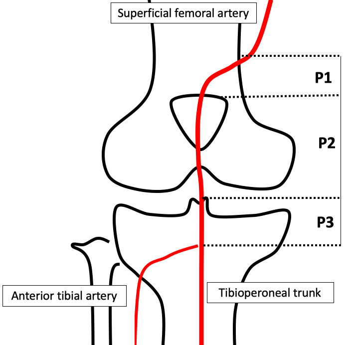
Figure 1: Zones of the popliteal artery
Regarding location, arterial lesions of the upper extremities are frequently affected in the brachial artery (40%), followed by the ulnar and radial arteries (25% in both cases), and the axillary artery in 30%. In the lower extremities, the most commonly affected is the deep femoral artery (37.2%), then the popliteal artery (30.7%), the crural artery (11%) and the common femoral artery (8.7%), with pseudoaneurysms representing 0.2% to 3.8%, with high amputation rates that can reach up to 12% (1,2,3,5). Doppler ultrasound has become the gold standard for visualizing pseudoaneurysms of peripheral arteries. The "yin-yang sign" is the common finding in most pseudoaneurysms; It is formed by the flow of blood from the arterial injury into the sac, creating turbulent flow as blood enters and exits with systole and diastole. (2,3,4)
Thus, the case presented here corresponds to an Ecuadorian patient with a popliteal artery pseudoaneurysm secondary to gunshot wounds. The diagnosis and treatment of these types of cases can be challenging, so we decided to share our experience regarding his clinical presentation and emergency management.
A 19-year-old Ecuadorian male resident in Quito with no significant medical history presented with bleeding in the popliteal fossa during the initial evaluation, which was controlled in an emergency setting using hemostasis and edge approximation. He was hemodynamically stable. He came to the hospital three months after suffering a gunshot wound to his left leg, presenting with increased pain in his leg and foot of moderate intensity. Physical examination revealed a visible and palpable mass in the left popliteal fossa, approximately 4 cm in diameter, pulsatile, non-mobile, with a soft consistency, and no pain on palpation.
Color Doppler revealed the presence of a pseudoaneurysm of the popliteal artery measuring 4.45 x 2.93 cm. Similarly to the angiotomography, an image was seen at the level of the distal third of the left thigh, 4.5 cm from the knee joint, in the femoral artery, a sac-like image was observed measuring 62 x 24 x 68 mm with an approximate volume of 53 cc, with the presence of an extensive mural thrombus and calcified atheromatous plaque (Figures 1 and 2).
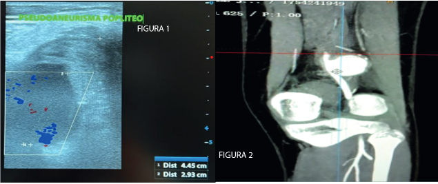
Figures 2 and 3: Doppler ultrasound and CT angiography of the femoral and popliteal arteries with the presence of post-traumatic pseudoaneurysm, respectively.
After the preliminary assessments, it was decided to take the patient to the operating room. The surgery consisted of resection of the aneurysm and primary revascularization with an autologous graft from the inverted great saphenous vein with end-to-end anastomosis (Figure 3).
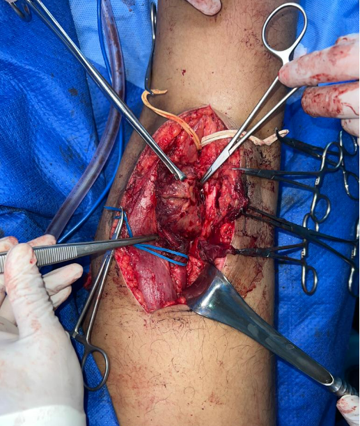
Figure 4: Resection of post-traumatic pseudoaneurysm.
The procedure was performed without complications. On the second day, the patient was in good health, with no fluid production at the drainage site, preserved motor strength, reflexes, and distal pulses. The decision was made to discharge him. He is currently in good health and performing his daily activities adequately.
Post-traumatic popliteal pseudoaneurysm is a common complication after traumatic injury to the popliteal region. (7,8,9) Most patients with this condition are male, with mean ages of 45.8 and 43.5 years, respectively. In our case, the gender is the same; however, it does not correspond to the usual age, as the patient was 19 years old. (8,9,10) Most pseudoaneurysms are caused by high or low energy trauma, such as car accidents, falls, and sports injuries. (9,10,11,12) In this case, the victim was a firearm. Patients may present with pain and tenderness in the popliteal region, as well as a pulsatile mass in this area. Additionally, they may experience symptoms of arterial insufficiency in the affected extremity, including intermittent claudication and decreased foot pulsation. (10,11)
Diagnosis is confirmed by imaging tests such as angiography, computed tomography (CT), and Doppler ultrasound, the methods used in the case described. Regarding treatment, noninvasive options include active surveillance and compression, while invasive options include embolization and surgery. (10,11,12,13)
Studies have shown that both angiography and CT are effective in detecting complications after endovascular or surgical treatment of popliteal pseudoaneurysm. Endovascular and surgical treatments are safe and effective options, with success rates of 89% and 97.2%, respectively. The complication rate is lower in endovascular treatment compared to surgical treatment; however, the success rate is significantly lower. Overall, the choice of treatment will depend on the individual circumstances of each patient; thus, in this case report, surgical resection of the pseudoaneurysm, along with autologous femoropopliteal bypass, was chosen without any surgical complications. In an additional study by Zhu et al., (8,10,11,12) a case of a patient with post-traumatic popliteal pseudoaneurysm presenting with a pulsatile mass in the popliteal region and leg pain is described. The authors used the same diagnostic and therapeutic methods as those used in our patient.
It is important to emphasize that prevention is key in the management of post-traumatic popliteal pseudoaneurysm; any activity that may increase the risk of traumatic injury to the popliteal region, such as contact sports or activities involving falls from height, should be avoided. Furthermore, patient education and awareness of the warning signs of the condition, as well as the importance of seeking immediate medical attention, are recommended. (9,10,11,12)
The main limitation of this case report was the lack of consideration of endovascular treatment; however, it is essential to note that this management is not available in the hospital unit where the patient was treated. Furthermore, a referral to a more complex hospital was not considered because the necessary material and human resources for its resolution were available, which are equally or more effective for this type of patient.
CONCLUSIONS
Popliteal artery pseudoaneurysm is a rare condition; however, its potential complications can compromise the patient's limb, so early identification, based on clinical and imaging studies, is of utmost importance. Timely surgical intervention can improve the patient's quality of life and allow for a more effective approach to managing vascular traumaappropriately . From the initial evaluation to the surgical procedure performed on each patient, proper identification of the area and anatomical location is essential to determine whether vascular compromise exists.
Funding: This clinical case was not conducted with any funding source.
Conflicts of interest: The authors declare that they have no conflicts of interest.
Authorization: The patient's signed informed consent is available.
Contribution statement:
MJ Bahamonde participated in data collection, design, editing, and final drafting of the manuscript.
MB Baño was the vascular surgeon who operated on the patient and obtained informed consent for publication.
DA Mora participated in data collection, contributed to the study design, edited, and finalized the drafting.
F. Pérez performed the diagnostic procedure and specific tasks in the final draft.
BIBLIOGRAPHIC REFERENCES
1. Melian CM, Giannopoulos S, Tsouknidas I, Volteas P, Virvilis D, Nicholson J, et al. Endovascular Repair of Popliteal Artery Injury Post-total Knee Arthroplasty is Safe and Effective: A Case Report and Systematic Review of the Literature. Ann Vasc Surg [Internet]. 2023 [cited June 16 2025];94:263–71. Available from: https://www.sciencedirect.com/science/article/abs/pii/S089050962300105X.
2. Fathima N. International Journal of Surgery Research Rare cases of pseudoaneurysm. 2019 [ cited June 16, 2025]; Available at: https://surgeryjournal.in/assets/archives/2022/vol4issue1/3-2-25-564.pdf.
3. Belai PM, Assis ZCB, Vieira TF de M, Vieira G dos SR, Almeida HA de, Lopes TV, et al. Post-traumatic pseudoaneurysm of the anterior tibial artery: therapeutic challenge. J Vasc Bras [Internet]. 2025 [cited June 16 2025];24:e20240120. Available at: https://www.scielo.br/j/jvb/a/FDKKnFCGw7spjKrQLLQzbVC/?lang=en.
4. Hübner CT, Vetter P, Heining SM, Pape HC, Hierholzer C. Traumatic pseudoaneurysm of the peroneal artery following lower extremity fracture – A case report and review of the literature. Trauma Case Rep [Internet]. 2025 [ cited June 16, 2025];56:101148. Available at: https://www.sciencedirect.com/science/article/pii/S2352644025000251.
5. Schwengber WK, Schnorr CC, Winckler GC, Paganella RB, Grudtner MA. Popliteal artery pseudoaneurysm of spontaneous occurrence : a case report . J Vasc Bras [Internet]. 2024 [cited June 16 2025];23:e20240021. Available at: https://www.scielo.br/j/jvb/a/wfv8HjFJzPWx8CvmQhbxqFq/?lang=en.
6. Monteleone N, Muratori F, Melani A, Schiavo A, Innocenti AA, Campanacci DA. A 54-Year-Old Man Who Developed a Femoral Pathologic Fracture from a Giant Popliteal Artery Pseudoaneurysm 7 Years After Ligation and Bypass of a Popliteal Artery Aneurysm: A Case Report and Literature Review. Am J Case Rep [Internet]. 2023 [cited June 16 2025];24:e937113-1. Available at: https://pmc.ncbi.nlm.nih.gov/articles/PMC9923776/.
7. Bodart E. Posterior tibial artery pseudoaneurysm: a rare complication following orthopedic surgery—a case report. J Surg Case Rep [Internet]. April 29, 2025 [cited June 16, 2025];2025(5). Available at: https://journals.aai.org/jscr/article/2025/5/rjaf288/8126588 DOI: https://dx.doi.org/10.1093/jscr/rjaf288.
8. Varothayan S, Vinojan S, Dhadchayini R, Gobinath S, Shathana P. Case report: Retrogenicular popliteal artery pseudoaneurysm following trivial knee hyperextension. Int J Surg Case Rep [Internet]. 2024 [cited June 16 2025];124:110439. Available at: https://www.sciencedirect.com/science/article/pii/S2210261224012203.
9. Rief M, Rief A, Bornemann- Cimenti H, Rief P. Idiopathic pseudoaneurysm of the popliteal artery with endovascular treatment: A case report. Radiol Case Rep [Internet]. 2023 [cited 16 Jun 2025];18(9):3336–40. Available from: https://www.sciencedirect.com/science/article/pii/S1930043323004338.
10. Alcala E, Manuel J, Romero G, Hugo P, Morales G, De D, et al. Gunshot-Induced Popliteal Artery Pseudoaneurysm: A Case Report. [Internet] 2025; [cited June 16 2025]: Available at: https://pubmed.ncbi.nlm.nih.gov/40642684/. DOI 10.7759/cureus.85660.
11. Rodríguez Martínez ADC, Barrientos-Villegas S, Palacios-Rodríguez PM, Fernando Hernández E, Martínez-Sosa IP, Valderrama-Treviño AI. Popliteal artery aneurysm in pediatric population: a review. International Surgery Journal. [Internet] 2024;11(8):1416–1419 Available from: https://www.ijsurgery.com/index.php/isj/article/view/10574.
12. Espinel C, Freire K, Conrado-Jiménez H, Camelo-Pardo G, Manrique-Hernández EF, Espinel C, et al. Hybrid management of recurrent post-traumatic pseudoaneurysm of the popliteal artery. Angiologia [Internet]. 2024 [cited 16 Jun 2025];76(5):334–338. Available from: https://scielo.isciii.es/scielo.php?script=sci_arttext&pid=S0003-31702024000500010&lng=es&nrm=iso&tlng=es.
13. Wang Y, Zheng H, Yao W, Ju S, Bai Y, Wang C, et al. Management of traumatic peripheral artery pseudoaneurysm: A 10-year experience at a single center. Journal of Interventional Medicine [Internet]. 2023 [ cited 16 Jun 2025];6(1):29–34. Available at : https://www.sciencedirect.com/science/article/pii/S2096360222000655.
14. EPOS&trade ; [Internet]. [cited June 16 2025]. Available at: https://epos.myesr.org/posterimage/esr/ecr2021/158861/mediagallery/897960?deliveroriginal=1.
Información adicional
redalyc-journal-id: 5728
