Image in focus
von Meyenburg Complex
 Manoj Gopal Madakshira
Tushar Pandey
Ashim Das
Manoj Gopal Madakshira
Tushar Pandey
Ashim Das
von Meyenburg Complex
Autopsy and Case Reports, vol. 9, no. 4, e2019107, 2019
Hospital Universitário da Universidade de São Paulo
Received: 05 July 2019
Accepted: 08 July 2019
A partial medical autopsy was carried out on a 50-year-old gentleman with a clinical diagnosis of acute severe necrotizing pancreatitis. The gross evaluation confirmed the presence of extensive necrotizing pancreatitis. However, the capsular and cut surface of the liver revealed the presence of subcapsular small bile stained lesions measuring 1 to 3 mm in diameter (Figure 1a, b). On histology, these bile stained lesions were seen to represent micro-hamartomas composed of a collection of dilated varying sized ducts lined by cuboidal epithelium in a fibrotic stroma. The cells lining these ducts were positive for Cytokeratin 7, confirming them to be of bile duct origin (Figure 1c, d). The subcapsular location of these lesions and characteristic histomorphology confirmed them to represent Von Meyenburg complexes (VMC).
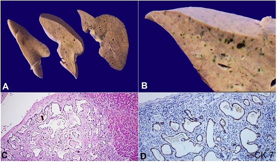
Figure 1
A and B - Cut surface of the liver show presence of multiple bile stained lesions measuring 3 to 15 mm in diameter in subcapsular location. B A closer view of the subcapsular bile stained lesions; C and D – Photomicrographs of the liver shows mis-shaped dilated bile ducts lined by cuboidal cells, embedded in a fibrotic stroma (H&E, 200X) and cuboidal cells lining the ducts show cytoplasmic staining for Cytokeratin 7.
VMC is a hamartomatous lesion and has been reported in 5.6% of autopsies.1 These micro-hamartomas are a part of the spectrum of ductal plate malformations which also include congenital hepatic fibrosis, Caroli’s disease, and adult polycystic disease.2 Embryonic ductal plate develops as a rounded structure between the primitive hepatocytes and portal mesenchyme.3 The ductal plate malformation is a result of an aberrant persistence of the embryonic ductal plate at varying levels of the biliary tree – smaller interlobular bile ducts are affected in VMC, while larger interlobular bile ducts are affected in Caroli’s disease. 2, 4 The defect lies at the stage of morphogenesis of bile duct formation which has its molecular basis in ciliogenesis and cellular polarization orchestrated by hepatocyte nuclear factors 1 and 6β.5
The importance of VMCs is in their simulation of neoplastic lesions. Utilizing the services of imaging and liver biopsy are essential to confirm the diagnosis of VMCs.6 There have also been anecdotal reports of malignant transformation of VMCs to cholangiocarcinoma. However, the low incidence of malignancy in case of VMCs suggest the importance played by secondary factors such as alcoholism, drugs, or infections in carcinogenesis.7
The authors retain an informed written consent for publication, and the manuscript is in accordance with the Institutional Ethics committee requirements.
References
Redston M, Wanless I. The hepatic von Meyenburg complex: prevalence and association with hepatic and renal cysts among 2843 autopsies [corrected]. Modern pathology: an official journal of the United States and Canadian Academy of Pathology. Inc. 1996;9(3):233-7.
Desmet VJ. Ludwig symposium on biliary disorders--part I. Pathogenesis of ductal plate abnormalities. Mayo Clin Proc. 1998;73(1):80-9. http://dx.doi.org/10.1016/S0025-6196(11)63624-0. PMid: 9443684
Terada T, Kitamura Y, Nakanuma Y. Normal and abnormal development of the human intrahepatic biliary system: a review. Tohoku J Exp Med. 1997;181(1):19-32. http://dx.doi.org/10.1620/tjem.181.19. PMid: 9149336
Awasthi A, Das A, Srinivasan R, Joshi K. Morphological and immunohistochemical analysis of ductal plate malformation: correlation with fetal liver. Histopathology. 2004;45(3):260-7. http://dx.doi.org/10.1111/j.1365-2559.2004.01945.x. PMid: 15330804
Raynaud P, Tate J, Callens C, et al. A classification of ductal plate malformations based on distinct pathogenic mechanisms of biliary dysmorphogenesis. Hepatology. 2011;53(6):1959-66. http://dx.doi.org/10.1002/hep.24292. PMid: 21391226
Venkatanarasimha N, Thomas R, Armstrong EM, Shirley JF, Fox BM, Jackson SA. Imaging features of ductal plate malformations in adults. Clin Radiol. 2011;66(11):1086-93. http://dx.doi.org/10.1016/j.crad.2011.05.008. PMid: 21840516
Jain D, Sarode VR, Abdul-Karim FW, Homer R, Robert ME. Evidence for the neoplastic transformation of Von-Meyenburg complexes. Am J Surg Pathol. 2000;24(8):1131-9. http://dx.doi.org/10.1097/00000478-200008000-00011. PMid: 10935654
Notes
Author notes
Correspondence Prof Ashim Das Post Graduate Institute of Medical Education and Research - Department of Histopathology, Research Block A, Sector 12, Chandigarh, India, Pin Code: 160012 Phone: +91 9872223744 ashim126@gmail.com
Conflict of interest declaration