Article / Clinical Case Reports
Oral ulcerative lesions in a post-liver-transplantation patient
Oral ulcerative lesions in a post-liver-transplantation patient
Autopsy and Case Reports, vol. 9, no. 1, e2018046, 2019
Hospital Universitário da Universidade de São Paulo
Received: 02 July 2018
Accepted: 15 August 2018
Abstract: Oral involvement is rarely found in histoplasmosis, except in its disseminated form, which is mostly observed in the severely immunocompromised host. Herein, we presented the case of a 36-year-old female with a previous history of liver transplant, who was hospitalized due to fever, chills, night sweats, diarrhea, and painful oral lesions over the last 3 days. The oral examination revealed the presence of painful shallow ulcers lined by a pseudomembrane in the gingiva and the soft and hard palate. The initial working diagnosis comprised cytomegalovirus reactivation or herpes simplex virus infection. The diagnostic work-up included incisional biopsies of the gingiva and the sigmoid colon. Both biopsies confirmed the diagnosis of histoplasmosis. Intravenous itraconazole was administered with significant improvement after 7 days. Although oral involvement is rare, histoplasmosis should be included in the differential diagnosis of oral lesions, particularly when the patient is immunosuppressed. This study reports a rare presentation of histoplasmosis involving the mucosa of the oral cavity and the colon.
Keywords: Histoplasmosis, Liver Transplantation, Oral ulcer, Immunosuppression.
INTRODUCTION
Histoplasmosis is an opportunistic fungal infection caused by Histoplasma capsulatum, a dimorphic fungus that lives in soil that is rich in birds and bats droppings.1 It is more prevalent in some regions in North, Central, and Latin America, as well as in Africa, and is typically found in tropical and temperate rural areas. The disease mostly occurs in the lungs and is acquired through the inhalation of dust particles from the soil contaminated with bat or bird excrement, which contains fungal spores—the infectious form of the microorganism.1
After reaching the airways, the microconidia are phagocytosed by the macrophages where they start to replicate. Hematogenous spread occurs within 2 weeks after infection. In immunocompetent patients, the macrophages have a fungicidal role by phagocytizing H. capsulatum, and therefore hamper the disease progression.1 However, in immunocompromised patients, such as those who are HIV-positive, transplant recipients, and patients with hematological neoplasm, the clinical course is more aggressive, with disseminated disease involving the lungs, skin, intestines, and oral mucosa.2 Here, we present an uncommon case of oral histoplasmosis, which was clinically misconceived as a viral infection.
CASE REPORT
A 36-year-old woman presented to the Emergency Department of the Cancer Center, complaining of fever, chills, night sweats, diarrhea, and painful oral lesions, over the past3 days. The patient had a medical history of liver transplantation8 years before, due to biliary cirrhosis, and was taking prednisone 5mg/day and mycophenolate sodium 360 mg. She also reported cytomegalovirus (CMV) infection 3 months after the transplantation, which was successfully treated with ganciclovir. The oral examination showed three well-defined shallow ulcerative lesions covered by fibrin with regular erythematous borders on the hard palate, measuring 1 cm (Figure 1A); on the vestibular and palatal gingiva between the 16 and 17 teeth measuring 0.5 cm (Figure 1B); and on the left soft palate near the uvula measuring 1.0 cm (Figure 1C).
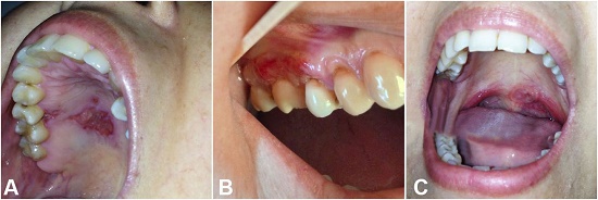
Figure 1
Gross examination of the oral lesions. A - Flat lesion covered with a fibrin pseudo membrane with regular erythematous borders on the hard palate; B - Ulcer on the vestibular and palatal gingiva between the 16 and 17 teeth; C - Ulcer on the left soft palate near the uvula.
According to the clinical findings and the previous history of CMV, the main diagnostic hypotheses were CMV reactivation or herpes simplex virus (HSV) infection. Exfoliative cytology of oral lesions was performed and an anti-CMV immunoglobulin (Ig)G test was requested. Because of the immunosuppression, ganciclovir 300mg tid was prescribed, but no improvement was observed within 7 days. Since the exfoliative cytology and the anti-CMV IgG tests were negative, the hypothesis of viral infection became less probable. A chest x-ray was normal, which ruled out lung involvement. Due to the worsening of oral lesions and the presence of gastrointestinal tract symptoms, two oral incisional biopsies—in the gingival and the palate—were taken, and a colonoscopy was performed with biopsy of a rectal lesion. The colonoscopy image showed areas with inflammatory process and eroded mucosa with an extensive ulcer measuring 2 cm on the descending colon extending to the rectum (Figure 2).
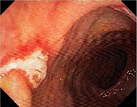
Figure 2
Colonoscopy image depicting area of inflammatory process with mucosal erosion and ulceration in the descending colon.
Histological sections of oral cavity biopsies revealed fragments of squamous mucosa with intense histiocytic inflammatory infiltrate associated with rounded fungal structures, which were consistent with H. capsulatum, and were confirmed by Gomori-Grocott’s staining in the gum and the palate (Figure 3). Similar findings were observed in the rectal biopsy. Based on the clinical and histopathologic findings, the final diagnosis was histoplasmosis (Figure 4).
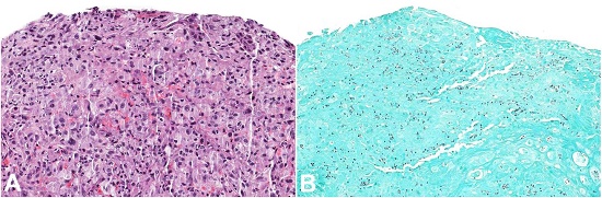
Figure 3
Photomicrograph of the oral biopsy. A - Areas of squamous mucosa with intense histiocytic inflammatory infiltrate associated with rounded fungal structures consistent with Histoplasma spp. (H&E 200X); B - Gomori-Grocott’s staining showing positivity for fungi (H&E 200X).
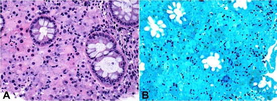
Figure 4
Photomicrograph of the rectal biopsy. A - Areas with intense histiocytic inflammatory infiltrate associated with rounded fungal structures consistent with Histoplasma spp. (H&E 400X); B - Gomori-Grocott staining showing positivity for fungi(400X).
The treatment consisted of itraconazole 300 mg/day with significant improvement of local and systemic symptoms after 7 days (Figure 5). The patient was followed up for 12 months with no further lesions.
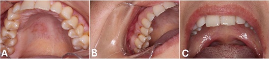
Figure 5
Oral examination after the treatment. Note the complete healing of the lesions on the hard palate (A), gingiva (B), and soft palate (C).
DISCUSSION
Clinical presentation of oral histoplasmosis is rare and the diagnosis is challenging. However, when present in the oral cavity, the involved sites include the tongue, the palate, the oral mucosa, the gingivae, and the pharynx. The mucosal involvement may occur as granular ulcerations, multiple painful ulcers and verrucous growths, as a deep ulcer surrounded by infiltrative edges with erythematous or white areas with irregular surfaces, as hardened and irregular nodular lesions accompanied by local lymphadenopathy, all of which mimic other infectious diseases or malignant tumors. This case report shows a case of histoplasmosis, which was initially misconceived as a viral infection.
The differential diagnoses of oral histoplasmosis include viral infections such as CMV and HSV. CMV is a ubiquitous herpes virus, which, depending on the studied population, infects 50%-100% ofhumans.3,4 Primary CMV infection in immune competent individuals presents most commonly without symptoms. However, in individuals with compromised immunity (e.g. liver transplant recipients) clinical disease may be fatal. CMV infection is the most common viral infection after solid transplant and usually appears during the first year after transplantation as observed in our case. The incidence after liver transplantation varies between 22% and 29%.5 CMV causes febrile illness, which is often accompanied by bone marrow suppression, and in some cases, involves other tissues including the transplanted liver allograft and gastrointestinal tract, causing abdominal pain and diarrhea. The skin and oral lesions usually present as chronic ulcers.3 Our patient presented fever, diarrhea, cutaneous rash, and oral lesions—features that are consistent with the diagnosis of CMV infection.
In contrast, HSV infection is a double-stranded DNA virus, which is usually acquired during childhood via infected saliva or direct contact with mucocutaneous lesions. After primary infection, the virus remains dormant until reactivation. The recurrent herpetic stomatitis is less common than the herpes labial and usually arises on keratinized surfaces.6 In immunocompromised patients, the recurrent HSV-1 infection may be atypical, with more extensive, slow-healing, and extremely painful lesions.7,8 In our case, the involvement of the keratinized areas, such as the palate and the gingiva, supported the hypothesis of the HSV infection.
The histopathological features of histoplasmosis are usually characteristic, but occasionally the organisms are scanty and not readily identified, which can preclude the correct diagnosis and consequently hamper the appropriate management.9 Fortunately, in the present case, the histopathologic examination results enabled a clear diagnosis of histoplasmosis.
Systemic antifungals are used to treat severe acute histoplasmosis as well as all chronic and disseminated cases. However, even with adequate treatment, the risk of failure and relapse does exist, so prolonged treatment is required.1 In the present case, the patient continues to be closely followed up to better evaluate the treatment response.
In conclusion, a case of histoplasmosis involving the oral cavity in an immunosuppressed patient is reported, which was initially misdiagnosed due to her previous medical history of CMV disseminated infection. The early and accurate diagnosis of histoplasmosis is essential for the correct treatment and cure.
REFERENCES
Souza BC, Munerato MC. Oral manifestation of histoplasmosis on the palate. An Bras Dermatol. 2017;92(5, Suppl 1):107-9. http://dx.doi.org/10.1590/abd1806-4841.20175751. PMid:29267463
Wheat LJ, Slama TG, Zeckel ML. Histoplasmosis in the acquired immune deficiency syndrome. Am J Med. 1985;78(2):203-10. http://dx.doi.org/10.1016/0002-9343(85)90427-9. PMid:3871588
Yadav SK, Saigal S, Choudhary NS, Saha S, Kumar N, Soin AS. Cytomegalovirus infection in liver transplant recipients: current approach to diagnosis and management. J Clin Exp Hepatol. 2017;7(2):144-51. http://dx.doi.org/10.1016/j.jceh.2017.05.011. PMid:28663679
López-Oliva MO, Flores J, Madero R, et al. Cytomegalovirus infection after kidney transplantation and long-term graft loss. Nefrologia. 2017;37(5):515-25. http://dx.doi.org/10.1016/j.nefro.2016.11.018. PMid:28946964
Simon DM, Levin S. Infectious complication soft solid organ transplantations. Infect Dis Clin North Am. 2001;15(2):521-49. http://dx.doi.org/10.1016/S0891-5520(05)70158-6. PMid:11447708
Arduino PG, Porter SR. Herpes simplex virustype 1 infection: overview onrelevantclinico-pathological features. J Oral Pathol Med. 2008;37(2):107-21. http://dx.doi.org/10.1111/j.1600-0714.2007.00586.x. PMid:18197856
Al-Dhafiri SA, Molinari R. Herpetic folliculitis. J Cutan Med Surg. 2002;6(1):19-22. http://dx.doi.org/10.1177/120347540200600104. PMid:11896419
Levitsky J, Duddempudi AT, Lakeman FD, et al. Detection and diagnosis of herpes simplex virus infection in adults with acute liver failure. Liver Transpl. 2008;14(10):1498-504. http://dx.doi.org/10.1002/lt.21567. PMid:18825709
Iqbal F, Schifter M, Coleman HG. Oral presentation of histoplasmosis in an immunocompetent patient: a diagnostic challenge. Aust Dent J. 2014;59(3):386-8. http://dx.doi.org/10.1111/adj.12187. PMid:24819556
Notes
Author notes
Correspondence Graziella Chagas Jaguar Stomatology Department - AC Camargo Câncer Center Rua Prof. Antônio Prudente, 211 - Liberdade - São Paulo/SP - Brazil CEP: 01509-900 Phone +55 (11) 2189-5129 graziellajaguar@gmail.com
Conflict of interest declaration