Original
Spirocerca lupi in dogs of Yucatan, Mexico: Case report and retrospective study
Spirocerca lupi en perros de Yucatán, México: Reporte de caso y estudio retrospectivo
Spirocerca lupi in dogs of Yucatan, Mexico: Case report and retrospective study
Revista MVZ Córdoba, vol. 24, no. 1, pp. 7145-7150, 2019
Universidad de Córdoba

This work is licensed under Creative Commons Attribution-NonCommercial-ShareAlike 4.0 International.
Received: 04 May 2018
Accepted: 04 June 2018
Published: 04 March 2019
Abstract: Objective. To describe the case report of a parasitized dog with Spirocera lupi in Yucatan, Mexico, as well as report cases registered in two laboratories during 18 years of parasitological and necropsy studies (2000-2017). Materials and methods. A case study is reported, with necropsy, histological and parasitological findings. Likewise, a retrospective study of cases reported in two laboratories where necropsies and faecal Flotation techniques (centrifugal and McMaster) were performed. Results. At the necropsy of the dog, three esophageal nodules were observed, which showed nematodes of S. lupi during the incision of the mass. The histological study showed an eosinophilic granuloma that contained the nematodes at its center, surrounded by a moderate inflammatory infiltrate formed by neutrophils, eosinophils, lymphocytes, plasma cells and macrophages, delimited by a capsule of fibrous connective tissue. In the retrospective study, prevalence of 0.18 and 0.48% were found by coprological tests and necropsy study, respectively. Conclusions. Spirocerca lupi is present in dogs from Yucatan, Mexico with low prevalence, producing in the esophagus of dogs lesions characterized by eosinophilic granulomas. The need to include this pathology in the differential diagnosis of esophageal and respiratory problems in dogs is disclosed.
Keywords: Coprological study, dog, eosinophilic granuloma, necropsy .
Resumen: Objetivo. Se describe el caso de un perro parasitado con Spirocera lupi en Yucatán, México, y además, se reportan los casos registrados en dos laboratorios durante 18 años de estudios parasitológicos y de necropsias (2000-2017). Materiales y métodos. Para el primer caso, se incluyen hallazgos de necropsia, histológicos y parasitológicos. Para los estudios retrospectivos se realizaron necropsias y estudios coprológicos de Flotación Centrifugada y de McMaster. Resultados. En el paciente del estudio de caso, durante la necropsia se observaron tres nódulos esofágicos que al realizar la incisión de los mismos, se visualizaron nematodos que correspondieron a S. lupi. En el estudio histológico se observó un granuloma eosinofílico que en su interior contenía el nematodo rodeado por un infiltrado inflamatorio moderado que estaba constituido por neutrófilos, eosinófilos, linfocitos, células plasmáticas y macrófagos; delimitado por una cápsula de tejido conectivo fibroso. En el estudio retrospectivo se encontraron prevalencias de 0.18 y 0.48% mediante pruebas coprológicas y estudios de necropsias, respectivamente. Conclusiones.Spirocerca lupi se encuentra presente en perros de Yucatán, México. Por lo tanto, sería importante considerar esta patología para el diagnóstico diferencial de problemas esofágicos y respiratorios en caninos.
Palabras clave: Estudio coprológico, granuloma eosinofílico, necropsia, perro .
INTRODUCTION
Spirocercosis is a disease caused by the nematode Spirocerca lupi (Rudolphi 1809). The dog is infected through the consumption of beetles, birds or small reptiles. The migratory larva causes hemorrhage, aortic stenosis, endarteritis, aneurysm or rupture of the aorta. Clinical signs of infection are regurgitation, vomiting, cough, dyspnea, weight loss and sudden death from damage to the aorta. Adult nematodes form nodules mainly in the esophagus, but occasionally they can occur in the wall of the stomach (1).
Due to larval migration, nodules or granulomas can form in other regions such as in thoracic organs, intestinal tract, urinary organs and in the connective tissue of the skin (2). Sarcomas can develop from the granulomas (3).
Spirocercosis is diagnosed by the history and clinical signs, coproparasitoscopic and molecular diagnosis, thoracic image, esophagoscopy and necropsy (4,5).
Spirocercosis caused by S. lupi occurs mainly in canines, and is predominant in tropical and subtropical areas. The majority of cases are reported in Israel, Italy, Greece, Turkey, India, Pakistan, Kenya and South Africa (6,7). In the American continent it has been reported in the United States, Brazil and Mexico (8,9).
In Querétaro, Mexico, S. lupi has been reported in dogs with a prevalence of 4.5% (9). In Yucatan, Mexico, Quiñones-Avila et al (10) were the first to report the presence of two dogs with S. lupi at the necropsy of 38 dogs (prevalence of 5.3%) and showed the importance of this parasite in dogs from the state of Yucatan.
Since this finding, in Yucatan, no case report of this nematode in dogs has been published. Therefore, the present study aims to describe the pathological case of a dog parasitized with S. lupi in Yucatan, as well as the report of cases in two laboratories during 18 years of parasitological and necropsy studies (2000-2017).
MATERIALS AND METHODS
Background. The report corresponds to a case study, with necropsy, histological and parasitological findings. As well as the retrospective study of cases reported in the Pathology and Veterinary Parasitology laboratories of the Campus of Biological and Agricultural Sciences of the Autonomous University of Yucatan (CCBA-UADY).
For the case study, a 12-year-old creole dog was received for a necropsy study. The dog had a history of regurgitation, vomiting and weight loss, was born in Merida, Yucatan, Mexico and never left the state of Yucatan. The state of Yucatan is located at a latitude of 19° 31 ‘- 21 ° 38› N and a longitude of 87° 22 ‘- 90 ° 25› W. The climate is tropical sub-humid with rains in summer. The maximum ambient temperature varies from 35 to 40 °C and the minimum from 10 to 16 ° C, with an average ambient temperature of 27 °C . The relative humidity varies from 65 to 90%, with an average of 80% and annual pluvial precipitation of 1,000 mm. Two annual seasons are presented: rain (from June to November) and dry season (from December to May) (11).
Diagnosis of the nematode and histopathological test. At the necropsy of the dog, three oesophageal nodules were observed. Incisions were made with the help of a scalpel blade to allow the exposure of the parasites. The parasitic nematodes were recovered alive and counted and identified with the help of a stereoscope microscope. The collected nematodes were maintained in 95% ethanol (12) and were identified according to Bowman et al (13).
Additionally, samples of the nodular lesions in the esophagus were collected and transferred to a wide-mouth plastic bottle with 10% buffered formalin with pH of 7.2, and maintaining the fixation sample ratio of 1:10. The vial was labeled and kept for 24 h for fixation.
The sample was processed using the paraffin embedding technique and Hematoxylin-Eosin staining. For this, the sample was dehydrated with different consecutive solutions of ethyl alcohol. They were then clarified with xylol and impregnated in paraffin until cooled. Once the paraffin was solidified and the cube formed, serial cuts of 5μm thickness were made, stained with Hematoxylin-Eosin and mounted with synthetic resin. Finally, the sample was revised with the aid of an optical microscope (14).
Retrospective study. To know the presence of cases of S. lupi in dogs in southeastern Mexico, the archives of the Parasitology and Veterinary Pathology laboratories of the CCBA-UADY were reviewed from January 2000 to December 2017. Additional information was also obtained from the positive cases such as the origin of the animals, age, breed and the excretion of eggs per gram of feces.
During the period of the retrospective study, 1,631 coprological studies of faecal samples from dogs were performed using the Centrifugal Flotation and McMaster techniques (12). The eggs of the nematodes were identified using the morphological descriptions and sizes described by Bowman et al (13). Also, in this period, 835 necropsies of dogs were performed according to the methodology described by Schueneman and Constantino (15).
RESULTS
During the necropsy of the dog, three nodules were observed in the esophagus (Figure 1), which showed reddish-colored adult nematodes during the incision of the nodular masses (Figure 2). The macroscopic lesions were located in the last third of the esophagus where each nodule had adherence to the wall of the esophagus, with one growing towards its lumen causing stenosis. In an esophageal region adjacent to a nodule, saccular dilatation was found. The nodules measured between 4 and 3.5 cm long, 2.5 and 3 cm wide and 2 and 3 cm high; and had firm to hard consistency. The nodules had a large central cavity, full of nematode parasites (reddish and whitish), surrounded by cell detritus and delimited by a fibrous connective tissue capsule that sometimes appeared calcified; in the three nodules a fistula was found that opened towards the lumen of the esophagus.
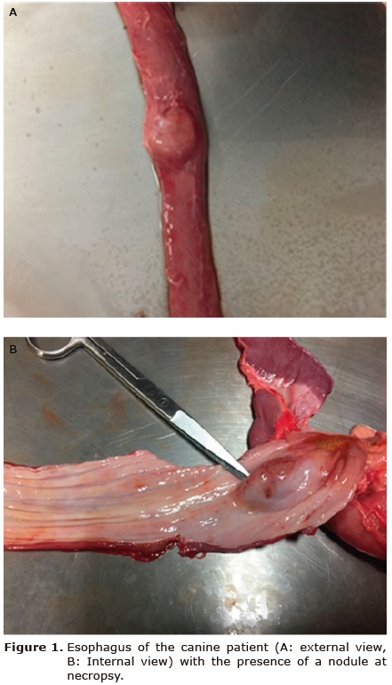
Figure 1
Figure 1. Esophagus of the canine patient (A: external view, B: Internal view) with the presence of a nodule at necropsy.
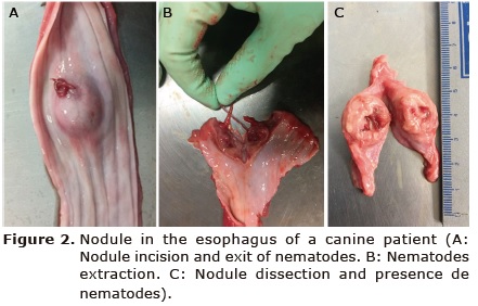
Figure 2
Figure 2. Nodule in the esophagus of a canine patient (A: Nodule incision and exit of nematodes. B: Nematodes extraction. C: Nodule dissection and presence de nematodes).
From the three nodules, 12 bright red adult nematodes were recovered, of which seven were males and five were females (Figure 3). The nematodes were classified as S. lupi (Figure 3). Males measured 4.2. ± 0.7 cm long and 1.0 ± 0.2 mm wide and females 7.0 ± 0.3 cm long and 1.5. ± 0.2 mm wide . The terminal part of the male presented in spiral form with lateral wing and papilla, as well as unequal spiracles. In the female, the vulva opens near the end of the esophagus and the uterus contains thick-covered eggs.
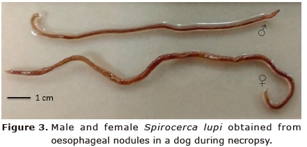
Figure 3
Figure 3. Male and female Spirocerca lupi obtained from oesophageal nodules in a dog during necropsy.
In the centrifugal flotation coprological study of the last case of the parasitology laboratory, S. lupi eggs were observed with a wide cover and with the presence of a larva with dimensions of 32.0 ± 1.8 μ of length x 12.2 ± 1.6 μ of width (Figure 4).
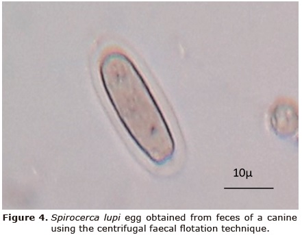
Figure 4
Figure 4.Spirocerca lupi egg obtained from feces of a canine using the centrifugal faecal flotation technique.
In the tissue sections esophageal nodule with eosinophilic granuloma were identified. In the central part of the nodule a parasitic structure of a nematode was identified, surrounded by a severe inflammatory infiltrate formed by neutrophils, eosinophils, lymphocytes, plasma cells and macrophages; which are delimited by a capsule of fibrous connective tissue formed by a large number of fibroblasts, fibrocytes and collagen fibers (Figure 5).
In the archives of the Veterinary Parasitology laboratory at the CCBA-UADY, 1631 coprological tests of dog feces samples were performed using the Centrifugal Flotation and McMaster techniques. Three cases of S. lupi were found, with a prevalence of 0.18% in feces. Also, the archives of the Animal Pathology laboratory at the CCBA-UADY from 2000 to 2017 were reviewed. In this period, 835 necropsies of canines were carried out and four dogs with oesophageal nodules containing adult nematodes of S. lupi were found, which correspond to a prevalence of 0.48%. All positive dogs reported in both laboratories came from the state of Yucatan, with no history of leaving the state.
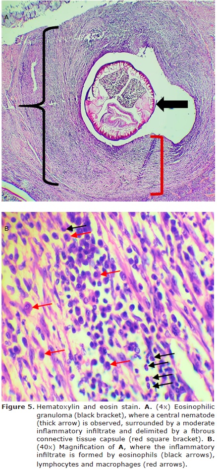
Figure 5
Figure 5. Hematoxylin and eosin stain. A. (4x) Eosinophilic granuloma (black bracket), where a central nematode (thick arrow) is observed, surrounded by a moderate inflammatory infiltrate and delimited by a fibrous connective tissue capsule (red square bracket). B. (40x) Magnification of A, where the inflammatory infiltrate is formed by eosinophils (black arrows), lymphocytes and macrophages (red arrows).
Table 1 shows the cases of dogs diagnosed with S. lupi in the Parasitology and Veterinary Pathology laboratories of the CCBA-UADY from 2000 to 2017.
| Diagnosis | Age of dog | Breed of dog | NNE* | EGF** |
| Case description | ||||
| Necropsy described in this study (Octuber 2017) | 12 years | Criollo | 12 | NA |
| Retrospective | ||||
| Coprologic (2002) | 5 years | Maltese | NA | 200 |
| Coprologic (2017) | 4 years | Criollo | NA | 150 |
| Necropsy (May 2000) | 5 years | Criollo | 9 | NA |
| Necropsy (Febraury 2014) | 7 years | Criollo | 7 | NA |
| Necropsy (Octuber 2017) | 9 years | Criollo | 8 | NA |
| NNE= Number of nematodes in esophagus; EGF=Eggs per gram of feces; * Diagnosed by necropsy, ** Diagnosed by McMaster technique, NA: Not applicable; | ||||
DISCUSSION
Spirocercosis has a worldwide distribution; however, is predominant in tropical and subtropical regions (6). The infection depends on the density of the dog population and the degree of interaction between the dog and the paratenic and intermediate hosts (mainly coprophagous beetles). The prevalence of this parasitic disease is variable and the majority of cases are reported in the Southeastern United States, Brazil, South Africa, Kenya and the Middle East (6,16,17).
Necropsy studies for the diagnosis of S. lupi in dogs have been carried out in different parts of the world, with prevalences of 14% in South Africa (16,18), 40.0% in Bangladesh (19), and 14.2% in Spain (20). In the present study, low prevalences of 0.18 and 0.48% were found, diagnosed in coprological tests and necropsies, respectively. These prevalences of S. lupi in dogs are also low compared to previous studies in Yucatan and Queretaro, where prevalences of 4.5% (9) and 5.3% (10) were found, respectively. This tendency to diminish the cases of S. lupi in dogs from Yucatan is probably associated to the constant use of anthelmintics and especially macrocyclic lactones in dogs (21), which have been shown to be effective to control this nematode (6,22).
Spirocercosis occurs mainly in dogs from rural areas or exposed to paratenic hosts. In Greece it was shown that dogs with owner presented lower prevalence (10%) of S. lupi compared with dogs that are used for hunting (21%) (23). In the present study it was not possible to investigate the origin of the dogs; however, most of the positive dogs were of criollo breed and adopted by their owners. This condition might favor exposure to the intermediate and paratenic hosts due to the habit of searching food in streets. In Yucatan, Mexico, different species of coprophagous beetles have been reported and the species Canthon leechi has preference for dog feces (24). In future studies it will be necessary to study the role of coprophagous beetles within the transmission cycle of S. lupi.
Different studies indicate that the age of dogs is not a factor associated with infection with S. lupi (6). However, in this study positive dogs for S. lupi were adult or geriatric animals aged 4 to 12 years.
Dogs become infected through the ingestion of L3 larvae of S. lupi. This occurs through the direct consumption of coprophagous beetles or the consumption of contaminated food; rodents, birds, rabbits and lizards as paratenic hosts are another potential transmission route. Larvae penetrate the stomach wall of dogs and migrate to the thoracic aorta via the gastric arteries. The larvae move to L4 and immature adults. In 3-4 months they migrate to the esophagus where they mature and are surrounded by granulomatous nodules as part of the dog’s inflammatory response (5,6).
During the necropsy of the dog three nodules were observed in the esophagus that during the incision of the mass allowed visualizing adult nematodes of S. lupi. In the histological study it was observed in the nodules with eosinophilic granuloma, with the presence of the nematode surrounded by a severe inflammatory infiltrate by neutrophils, eosinophils, lymphocytes, plasma cells and macrophages. These findings are in agreement with that reported by Diakou et al (25) who describe that the granulomas consist of a central area that are surrounded by degenerate and viable eosonophils, as well as the infiltration of neutrophils as part of the animal’s immune response. Likewise, these are delimited by connective tissue infiltrated predominantly by mononuclear cells, specifically histiocytes, plasma cells and lymphocytes.
The pathogenesis of tumor induction by S. lupi has not been well elucidated (26,27). It is suggested that oesophageal sarcomas are produced by S. lupi larvae (3). It is believed that parasites interfere with cellular processes of relevance in carcinogenesis, acting in intercellular communication or through the secretion of molecules with carcinogenic potential (28). Fibrosarcomas and osteosarcoma can occur with the possibility of metastasis in the lungs (3), a pathology that did not occur in the case described in this study. The development of these tumors leads to oesophageal obstructions and dyspnea, which can lead to a severe clinical outcome that can lead to the death of dogs.
It is concluded that S. lupi is present in dogs from Yucatan, Mexico at low prevalence, producing lesions characterized by eosinophilic granulomas in the esophagus of dogs. The need to include this pathology in the differential diagnosis of esophageal and respiratory problems in dogs is revealed.
Interest conflict.
The authors declare no conflict of interests of any kind.
REFERENCES
1. Psáder R, Balogh M, Pápa K, Sterczer A, Lukács Z, Harnos A. Occurrence of Spirocerca lupi infection in hungarian dogs referred for gastroscopy. Parasitol Res. 2017; 116:S99–S108. https://doi.org/10.1007/s00436-017-5496-9
2. Lobetti R. Spirocercosis. Canine and feline gastroenterology. Washabau RJ, Day MJ (ed). Elsevier Saunders; St. Luis: 2013.
3. Ranen E, Lavy E, Aizenberg I, Perl A., Harrus S. Spirocercosis - associated esophageal sarcomas in dogs. A retrospective study of 17 cases (1997-2003). Vet Parasitol. 2004; 119(2-3):209-221. https://doi.org/10.1016/j.vetpar.2003.10.023
4. Christie J, Schwan EV, Bodenstein LL, Sommerville JE, Van der Merwe LL. The sensitivity of direct faecal examination, direct faecal flotation, modified centrifugal faecal flotation and centrifugal sedimentation/flotation in the diagnosis of canine spirocercosis. J S Afr Vet Assoc. 2011; 82:71-75.
5. Wright I. Spirocerca lupi - a potential future problem for pets travelling to Europe. Companion Anim. 2015; 20(3):156-160. https://doi.org/10.12968/coan.2015.20.3.156
6. van der Merwe LL, Kirbergera RM, Clift S, Williams M, Keller N, Naidoo V. Spirocerca lupi infection in the dog: A review. Vet J. 2008; 176:294-309. https://doi.org/10.1016/j.tvjl.2007.02.032
7. Giannelli L, Baldassarre V, Ramos RAN, Lia RP, Furlanello T, Trotta M et al. Spirocerca lupi infection in a dog from southern Italy: an old fashioned disease? Parasitol Res. 2014; 113:2391. https://doi.org/10.1007/s00436-014-3912-y
8. Elias F, Barros RM, Santos-Junior HL, Eloi RSA, Silva V, Freitas F, Fonseca-Alves CE. Pathological alterations in dogs resulting from parasitism by Spirocerca lupi. Acta Sci Vet. 2016; 44(Suppl 1):145. http://revistas.bvs-vet.org.br/actascivet/article/view/31571/34927
9. Cantó GJ, García MP, García A, Guerrero MJ, Mosqueda J. The prevalence and abundance of helminth parasites in stray dogs from the city of Queretaro in central Mexico. J Helminthol. 2011; 85:263-269. https://doi.org/10.1017/S0022149X10000544
10. Quiñones-Avila F, Espaine AL, Rodríguez-Vivas RI, Domínguez-Alpizar J. Contribución al estudio de los helmintos del tracto digestivo en perros de la ciudad de Mérida, Yucatán, México. Rev AMMVEPE 1998; 9(6):191-193.
11. INEGI (Instituto Nacional de Estadística, Geografía e Información) [Base de datos de internet]. Anuario estadístico y geográfico de Yucatán México. 2017 [actualizado el 28 de abril de 2018]. URL Disponible en: http://www.datatur.sectur.gob.mx/ITxEF_Docs/YUC_ANUARIO_PDF.pdf
12. Rodríguez-Vivas RI editor. Técnicas para el diagnóstico de parásitos con importancia en salud pública y veterinaria. AMPAVE-CONASA; México DF: 2015.
13. Bowman DD, Lynn RC, Eberhard ML. Georgi’s parasitology for veterinarians. 8th edn. St. Louis, Missouri; Saunders: 2003.
14. Mescher AL. Junqueira’s Basic Histology: Text and Atlas. 14th Edition. Ed. McGraw-Hill Education; New York, USA: 2016.
15. Schueneman de AA, Constantino CF. Técnicas de necropsia en animales domésticos. 2a edición. Manual Moderno; México, D.F: 2002.
16. Minnaar WN, Krecek RC. Helminths in dogs belonging to people in a resource-limited urban community in Gauteng, South Africa. J Vet Res. 2001; 68:111–117. PMID: 11585088
17. Dantas-Torres F, Otranto D. Dogs, cats, parasites, and humans in Brazil: opening the black box. Parasit Vectors. 2014; 7:22. https://doi.org/10.1186/1756-3305-7-22
18. Lobetti R. Follow-up survey of the prevalence, diagnosis, clinical manifestations and treatment of Spirocerca lupi in South Africa. J S Afr Vet Assoc. 2014; 85(1):7. https://doi.org/10.4102/jsava.v85i1.1169
19. Shubhagata D, Abdul A, Mohammad MH, Suchandan S, Muraduzzaman M. Spirocercosis in stray dogs of Chittagong Metropolitan area of Bangladesh: an epidemiological and pathological investigation. Vet World. 2011; 4:485-491. https://doi.org/10.5455/vetworld.2011.485-491
20. Chikweto A, Bhaiyat MI, Tiwari KP, de Allie C, Sharma RN. Spirocercosis in owned and stray dogs in Grenada. Vet Parasitol. 190; 2012:613-616. https://doi.org/10.1016/j.vetpar.2012.07.006
21. Rodriguez-Vivas RI, Ojeda-Chi MM, Trinidad-Martinez I, Pérez de León AA. First documentation of ivermectin resistance in Rhipicephalus sanguineus sensu lato (Acari: Ixodidae). Vet Parasitol. 2017; 233:9-13. https://doi.org/10.1016/j.vetpar.2016.11.015
22. Austin CM, Kok DJ, Crafford D, Schaper R. The efficacy of a topically applied imidacloprid 10 %/moxidectin 2.5 % formulation (Advocate®, Advantage® Multi, Bayer) against immature and adult Spirocerca lupi worms in experimentally infected dogs. Parasitol Res. 2013; 112:91–108. https://doi.org/10.1007/s00436-013-3284-8
23. Mylonakis ME, Koutinas AF, Liapi MV, Saridomichelakis MN, Rallis TS. A comparison of the prevalence of Spirocerca lupi in three groups of dogs with different life and hunting styles. J Helminthol. 2001; 75:359-361. PMID: 11818054
24. Rodríguez-Vivas RI, Basto-Estrella G, Reyes-Novelo E, Delfín-González H, Pérez-Cogollo LC, Ojeda-Chi M et al. Escarabajos estercoleros en Yucatán, México: Especies, distribución estacional e incorporación de excretas al suelo. Rev Nuestro Agro. 2014; 1:18-24.
25. Diakou A, Karamanavi E, Eberhard M, Kaldrimidou E. First report of Spirocerca lupi infection in red fox Vulpes vulpes in Greece. Wildl Biol. 2012; 18(3):333-336. https://doi.org/10.2981/11-094
26. Mukorera V, van der Merwe LL, Lavy E, Aroch I, Dvir E. Serum alkaline phosphatase activity is not a marker for neoplastic transformation of esophageal nodules in canine spirocercosis. Vet Clin Pathol. 2011; 40(3):389-392. https://doi.org/10.1111/j.1939-165X.2011.00331.x
27. Klainbart S, Mazaki-Tovi M, Auerbach N, Aizenberg I, Bruchim Y, Dank G. Spirocercosis - associated pyothorax in dogs. Vet J. 2007; 173(1):209-214. https://doi.org/10.1016/j.tvjl.2005.08.019
28. Santos ASO, Silveira LS, Lemos LS, Moreira L, Silva AC, Carvalho ECQ. Aneurismas aórticos pela espirocercose canina em Brasília (Relato de caso). Rev Bras Saúde Prod Anim. 2004; 5(1):25-30. http://revistas.ufba.br/index.php/rbspa/article/view/628/385