Comunicaciones Breves
Double Renal Artery in Cerdocyon thous
Arteria renal doble en Cerdocyon thous
 Shirley Viana Peçanha shipeviana@gmail.com
Raquel Batista Junger de Carvalho raqbatista@gmail.com
Carlos Augusto Santos-Sousa augusto.anato@gmail.com
Emanoelli Aparecida Rodrigues dos Santos aleksandro_ss@yahoo.com.br
Paulo Souza-Junior paulosouza@unipampa.edu.br
Marcelo Abidu-Figueiredo marceloabidu@gmail.com
Shirley Viana Peçanha shipeviana@gmail.com
Raquel Batista Junger de Carvalho raqbatista@gmail.com
Carlos Augusto Santos-Sousa augusto.anato@gmail.com
Emanoelli Aparecida Rodrigues dos Santos aleksandro_ss@yahoo.com.br
Paulo Souza-Junior paulosouza@unipampa.edu.br
Marcelo Abidu-Figueiredo marceloabidu@gmail.com
Double Renal Artery in Cerdocyon thous
Revista MVZ Córdoba, vol. 25, no. 1, p. 1713, 2020
Universidad de Córdoba
Received: 08 July 2019
Accepted: 09 September 2019
Published: 03 February 2020
Abstract: Objective. The aim of this study was explored the duplicity of renal artery in a specimen of Cerdocyonthous, focusing on the possibilities of clinical-surgical implication of this anatomical variation. Materials and methods. Were dissected 32 specimens of Cerdocyon thous, obtained from the collections of the Laboratório de Ensino e Pesquisa em Morfologia dos Animais Domésticos e Selvagens do Departamento de Anatomia Animal e Humana, da Universidade Federal Rural do Rio de Janeiro e Laboratório de Anatomia Animal da Universidade Federal do Pampa. Results. Were observed a numerical variation in the left renal artery in an adult female cadaver. The left kidney had two renal arteries, one cranial and another caudal. The first renal artery of the left kidney, measuring 2.25 cm in length, originated laterally from the abdominal aorta at the level of the third lumbar vertebra. Moreover, it emanated two pre-hilar branches, one dorsal and one ventral, with the ventral branch supplying also to the adrenal gland. The second renal artery also originated laterally from the abdominal aorta at the level of the third lumbar vertebra and measured 2.36 cm in length. It also emitted two pre-hilar branches, one cranial and another caudal, which emitted the ureteral branch. Conclusions. Numerical variations of the renal arteries should be considered in the execution of surgical, radiological and experimental procedures in order to avoid mistakes made due to lack of knowledge of the possibility these variations both in domestic and wild animals.
Keywords: Anatomical variation, crab-eating-fox, urogenital system, wild carnivores .
Resumen: Objetivo. El objetivo de este estudio fue explorar la duplicidad de la arteria renal en un espécimen de Cerdocyon thous y centrarse en las posibilidades de la implicación clínico-quirúrgica de esta variación anatómica. Materiales y Métodos. Fueron disecados 32 especímenes de Cerdocyon thous, obtenidos de las colecciones del Laboratorio de Ensayo y Pesquisa en Morfología de Animales Domésticos y Salvajes del Departamento de Anatomía Animal y Humana, de la Universidad Federal Rural del Río de Janeiro y del Laboratorio de Anatomía Animal de la Universidad Federal del pampa. Resultados. Fue observado una variación numérica en la arteria renal izquierda en un cadáver hembra adulto. El riñón izquierdo tenía dos arterias renales, una craneal y otra caudal. La primera arteria renal del riñón izquierdo, que mide 2,25 cm de extensión se originó lateralmente desde la aorta abdominal a nivel de la tercera vértebra lumbar. Además, emanaba dos ramas prehiliares, una dorsal y otra ventral, con la rama ventral que se suministra también a la glándula suprarrenal. La segunda arteria renal también se originó lateralmente desde la aorta abdominal a nivel de la tercera vértebra lumbar y midió 2,36 cm de extensión. También emitió dos ramas prehiliares, una craneal y otra caudal, que emitieron la rama uretral. Conclusiones. Deben considerarse las variaciones numéricas de las arterias renales en la ejecución de procedimientos quirúrgicos, radiológicos y experimentales para evitar errores debido a la falta de conocimiento de la posibilidad de estas variaciones tanto en animales domésticos como salvajes.
Palabras clave: Variación anatómica, zorro perruno, sistema urogenital, carnívoros silvestres. .
INTRODUCTION
Cerdocyon thous is a neotropical wild canid that possesses an omnivorous diet comprising of small vertebrates, invertebrates and fruits (1,2,3,4). C. thous exhibits robust dimensions - 1meter length and 3.7 - 11 kg weight. It is a short, stout animal with a predominantly gray and yellow colored coat and a dark band on its back. Its habitat ranges from closed forests to open fields, occurring throughout the Brazilian territory except for the Amazon forest (3).
The knowledge about variations in renal vascular anatomy has great applicability in the semiotic and surgical exploration of kidneys (5,6). In mammals, the right and left renal arteries, which originate on each lateral side of the abdominal aorta, irrigate the respective kidneys (7).
The renal arteries transport approximately 20% of the arterial blood of the abdominal aorta (7). Usually the right renal artery is more cranial than the left, in accordance with the relatively more cranial position of the right kidney in most species (7).
Numeric variations in renal arteries have been previously described in dogs (8,9), goats (10) and rabbits (11). These anatomical variations are occasionally identified in radiological and routine dissection studies.
However, with respect to morphology, there are a few studies in these species, which have shown that it is important to obtain anatomical information of the animals both for future works and for the preservation of the species, in captivity as well as natural habitat (12). In this study, we explored the duplicity of renal artery in a specimen of Cerdocyon thous, focusing on the possibilities of clinical-surgical implication of this anatomical variation.
MATERIALS AND METHODS
Were dissected 32 specimens of Cerdocyon thous, obtained from the collections of the Laboratório de Ensino e Pesquisa em Morfologia dos Animais Domésticos e Selvagens (LEPeMADS) do Departamento de Anatomia Animal e Humana, da Universidade Federal Rural do Rio de Janeiro e Laboratório de Anatomia Animal da Universidade Federal do Pampa. We observed a numerical variation in the left renal artery in an adult female cadaver that was collected from the highway in Serra dos Órgãos National Park (license: ICMBio / SISBIO 33667). The cadaver was identified and positioned in right lateral decubitus.
For fixation of the specimen, the thorax was first opened and dissected and a urethral catheter number 6 was introduced in its thoracic aorta. The arterial system was then “washed” with 0.9% NaCl solution, and fixed with 10% formalin solution. Next, aqueous solution (1:1 dilution) of Petrolátex S-65 (Refinery Duque de Caxias-REDUC- Petrobras, Duque de Caxias-RJ) was injected together with a dye, Suvinil xadrez.. Finally, the cadaver was immersed in a 50 L low density polyethylene box containing 10% formaldehyde solution to complete the latex fixation and polymerization process. After 7 days of latex injection, the cadaver was washed in running water, and its abdominal cavity was opened and dissected to demonstrate the kidneys, their respective vessels and skeletopy. Using a digital caliper (0–150 mm, 0.01 mm resolution, accuracy ±0.02 mm, Eda.), renal measurements (length, width and thickness) and length of the renal arteries were obtained.
RESULTS
We observed that the specimen had a 64.1 cm cranio-sacral length. Its right kidney was 5.11 cm long, 1.75 cm wide and 2.57 cm thick, and was located between the first and the third lumbar vertebrae (L1 and L4). In the right kidney, there was only one renal artery, which was 2.01 cm in length. On the other hand, the left kidney was 5.27 cm long, 2.24 cm wide and 1.77 cm thick, and was located between the third and the fifth lumbar vertebrae (L3 and L5). Unlike the right kidney, the left kidney had two renal arteries (Figure 1), one cranial (1) and another caudal (2).
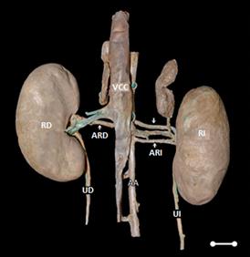
Figure 1
Photomacrography showing the double left renal artery in C thous CVCcaudal vena cava AAabdominal aorta RKright kidney LKleft kidney RRAright renal artery LRAleft renal artery ↓ cranial left renal artery ↑ caudal left renal artery RUright ureter LUleft ureter Scale bar 1cm
The first renal artery of the left kidney, measuring 2.25 cm in length, originated laterally from the abdominal aorta at the level of the third lumbar vertebra (L3). Moreover, it emanated two pre-hilar branches (Figure 2), one dorsal and one ventral, with the ventral branch supplying also to the adrenal gland. The second renal artery also originated laterally from the abdominal aorta at the level of the third lumbar vertebra (L3) and measured 2.36 cm in length. This artery also emitted two pre-hilar branches, one cranial and another caudal, which emitted the ureteral branch.
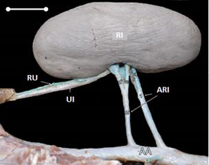
Figure 2
Photomacrography of the lateral view showing two pre-hilar branches of the each left renal artery in C. thous: AA-abdominal aorta, LK-left kidney, LU-left ureter, UB-ureteral branch, LRA-left renal artery (1-cranial LRA: a-dorsal pre-hilar branch, b-ventral pre-hilar branch; 2-caudal LRA: c-cranial pré-hilar branch, d-caudal pre-hilar branch). Scale bar 1cm.
DISCUSSION
Numerical variations of renal arteries have been described in different animals. The frequency of double renal arteries in dogs has been shown to be 12.80% (64 from 500 dogs) (8) and 9.72% (14 from 144 dogs) (9). In cats (13) and rabbits (11) only case reports were made, however their descriptions in wild animals remain scarce. In the case of C. thous, 64 kidneys were inspected and the occurrence of duplicity in renal arteries was recorded only in the left kidney of one specimen, thereby corresponding to 1.6% of the cases.
In humans, variations in the arrangement and distribution of renal arteries have also been studied by SAMPAIO and PASSOS (6), who cited cases of duplication (7.9%), triplication (1.9%), and even the occurrence of arterial branches supplying both the upper pole and the lower pole of the kidney. In these cases, the authors have suggested the use of the term “multiple renal arteries” because they are normal terminal segments without anastomoses between them and, from the physiological point of view, are important for renal blood supply. The same authors reinforced the importance of adequacy in the nomenclature used for such vessels and discouraged the usage of terms “aberrant,” “accessory,” “extra,” and “supernumerary,” since they convey the false idea of being elements with little functional importance.
In humans, the presence of multiple renal arteries increases the complexity of kidney transplantation (14), as well as correlates with renal pathologies, when compared to organs that are supplied by a single renal artery (6). The complexity of the vascular variations can also modify the technical possibilities of the surgical procedure and, according to Khamanarong et al (14) a complete understanding of renal vascular anatomy provides efficiency and safety in surgical and radiological procedures. Therefore, any abdominal surgery that requires mobilization or hemostatic control of the renal vessels requires a systematic search for possible vascular anatomical variations.
In domestic dogs, the renal artery usually has a pre-hilar bifurcation into the dorsal and ventral branches (15). In this case report, we could show that the cranial artery had this bifurcation pattern, where the artery bifurcated into a cranial and a caudal branch. The length of the two arteries was similar, however visually the caliber of the more cranially positioned artery was slightly larger than the flow. Both seemed functionally relevant. Altogether, the anatomical variation corresponds to the renal artery with caudal origin, which then emanated into the ureteral branch.
The disclosure about findings of anatomical variations is important for veterinary medical practice involving domestic and wild animals and should be performed as a way of raising awareness among professionals, contributing to the success and improvement of different clinical and surgical protocols.
Due to a wide distribution in the Brazilian territory, hundreds of Cerdocyonthous specimens are sent annually to rehabilitation centers and zoos in Brazil. These also include animals that are victims of poly-trauma by hunting, trampling and fire or that have been debilitated by various diseases (16,17). The incidence of abdominal pathologies is so high that a Doppler sonographic study was performed to characterize the dimensions of renal arteries (18) in the context of urological conditions such as azotemia (19) and renal trauma resulting in nephrectomy (20).
Therefore, numerical variations of the renal arteries should be considered in the execution of surgical, radiological and experimental procedures in order to avoid mistakes made due to lack of knowledge of the possibility of such variations both in domestic and wild animals.
Conflict of interests
The authors declare that there are no potential conflicts of interest regarding the research, authorship or publication of this article.
REFERENCES
1. Machado FA, Hingst-Zaher E. Investigating South American biogeographic history using patterns of skull shape variation on Cerdocyon thous (Mammalia: Canidae). Biol J Linn Soc. 2009; 98(1):77-84. https://doi.org/10.1111/j.1095-8312.2009.01274.x
2. Perini FA, Russo CAM, Schrago CG. The evolution of South American endemic canids: a history of rapid diversification and morphological parallelism. J Evol Biol. 2010; 23(2):311-322. https://doi.org/10.1111/j.1420-9101.2009.01901.x
3. Trigo TC, Rodrigues MLF, Kasper CB. Carnívoros Continentais. In: Weber MM, Roman C, Cáceres NC. Mamíferos do Rio Grande do Sul. Santa Maria: UFSM; 2013. https://editoraufsm.com.br/mamiferos-do-rio-grande-do-sul
4. Kasper CB, Trinca CS, Sanfelice D, Mazi FD, Trigo TC. Os Carnívoros. In: Gonçalves GL, Quintela FM, Freitas TRO. (eds.) Mamíferos do Rio Grande do Sul. Porto Alegre: Pacartes; 2014. https://www.catarse.me/mamiferosrs
5. Sampaio FJ, Aragão AH. Anatomical relationship between the intrarenal arteries and the kidney collecting system. J Urol 1990; 143(4):679-681. https://doi.org/10.1016/s0022-5347(17)40056-5
6. Sampaio FJB, Passos MARF. Renal arteries: anatomic study for surgical and radiological practice. Surg Radiol Anat 1992; 14(2):113-117. https://link.springer.com/article/10.1007/BF01794885
7. König HE, Liebich HG. Anatomia dos animais domésticos: texto e atlas colorido. 6th ed. Porto Alegre: Artmed; 2016.
8. Reis RH, Tepe P. Variation in the pattern of renal vessels and their relation to the type of posterior vena vava in the dog (Canis familiaris). Am. J. Anat 1956; 99:1-15. https://doi.org/10.1002/aja.1000990102
9. Sajjarengpong K, Adirektaworn A. The variations and patterns of renal arteries in dogs. The Thai Journal of Veterinary Medicine 2006; 36(1):39-46. www.tci-thaijo.org/index.php/tjvm/article/view/36332
10. Abidu-Figueiredo M, Roza MS, Passos NC, Silva BX, Scherer PO. Artéria renal com dupla origem na porção abdominal da aorta em caprino. Acta Veterinaria Brasilica 2009; 3(1):38-42. https://doi.org/10.21708/avb.2009.3.1.970
11. Almeida BB, Barreto UH, Costa OM, Abidu-Figueiredo M. Double renal artery in rabbits. Biosci J 2013; 29(5):1294-1295. http://www.seer.ufu.br/index.php/biosciencejournal/article/view/22287
12. Machado LC, Roballo KCS, Cury FS, Ambrósio CE. Female repro-ductive system morphology of crab-eating fox (Cerdocyon thous) and cryopreservation of genetic material for animal germplasm bank en-richment. Anat Histol Embryol 2017; 46(6): 539-546. http://dx.doi.org/10.1111/ahe.12306
13. Pestana FM, Roza MS, Hernandez JMF, Silva BX, Abidu-Figueiredo M. Artéria renal dupla em gato: relato de caso. Semina: Cien Agrar 2011; 32(1):325-330. http://dx.doi.org/10.5433/1679-0359.2011v32n1p327
14. Khamanarong K, Prachaney P, Utraravichien A, Tong-Un T, Sri-paoraya K. Anatomy of renal arterial supply. Clin Anat 2004; 17:334-336. https://doi.org/10.1002/ca.10236
15. Evans, H. E., A. Delahunta. Miller’s Anatomy of the Dog. 4th ed. St Louis (MO): Saunders Elsevier; 2013. https://www.elsevier.com/books/millers-anatomy-of-the-dog/evans/978-1-4377-0812-7
16. Tavares DS, Varjão COV, Santos AH, Iamagute LS, Conceição AM, Barros SLB. Amputação de membro pélvico de cachorro-do-mato (Cerdocyon thous) devido à osteomielite pós cirurgia de correção de fratura: relato de caso. Rev Educ Cont Vet Med Zootec 2013; 11(3):98. https://www.revistamvez- crmvsp.com.br/index.php/recmvz/article/view/21397
17. Sposito GC, Gorios A, Camargo LP, Campos MAR, Estrella JPN, Credie LFGA, et al. Anestesia epidural sacrococcígea em osteossíntese femoral em cachorro-do-mato (Cerdocyon thous). Relato de caso. Rev Educ Cont Vet Med Zootec 2016; 14(2):46. https://www.revistamvez-crmvsp.com.br/index.php/recmvz/article/view/31823
18. Silva ASL, Feliciano MAR, Motheo TF, Oliveira JP, Kawanami AE, Werther K, et al. Mode B ultrasonography and abdominal Doppler in crab-eating-foxes (Cerdocyon thous). Pesq Vet Bras 2014; 34(Suppl 1):23-28. https://doi.org/10.1590/S0100-736X2014001300005
19. Silva TR, Nogueira AFS, Santana AE. Avaliação dos perfis bio-químicos hepático e renal em Cerdocyon thous (Cachorro do mato) Sororreagentes a Leptospira spp. no território brasileiro. Ars Veteri-naria 2013; 29(4):9. http://dx.doi.org/10.15361/2175-0106.2013v29n4p9
20. Piccoli RJ, Thomazoni D, Druziani JT, Hamamura M, Carvalho AL. Nefrectomia total unilateral em cachorro-do-mato (Cerdocyon thous). Acta Sci Vet 2017; 45(Suppl 1):228. http://www.ufrgs.br/actavet/45-suple-1/CR_228.pdf
Additional information
How to cite (Vancouver).: Peçanha SV, Junger de CRB, Santos-Sousa CA, Rodrigues SEA, Souza-Junior P, Abidu-Figueiredo M. Double Renal Artery in Cerdocyon thous. Rev MVZ Cordoba. 2020; 25(1):e1713. DOI: https://doi.org/10.21897/rmvz.1713