Review Article
Development of anthropomorphic computational phantoms at the UFPE
Development of anthropomorphic computational phantoms at the UFPE
Brazilian Journal of Radiation Sciences, vol. 11, no. 1, e2243, 2023
Sociedade Brasileira de Proteção Radiológica
Received: 30 December 2022
Accepted: 03 January 2023
Published: 09 March 2023
Abstract: To evaluate the amount of energy deposited in radiosensitive organs and tissues of the human body, when an anthropomorphic phantom is irradiated, researchers in numerical dosimetry use the so-called exposure computational models (ECMs). One can imagine an ECM as a virtual scene composed of a phantom in a mathematically defined position in relation to a radioactive source. The source in these ECMs produces the initial state of the simulation: the position, direction, and energy with which each particle enters the phantom are essential variables. For subsequent states of a particle history, robust Monte Carlo (MC) codes are used. For the subsequent states of a particle's history, robust Monte Carlo (MC) codes are used, which simulate the average free path that the particle performs without interacting, its interaction with the atoms in the medium and the amount of energy deposited per interaction. MC codes also evaluate normalization quantities, so the results are printed in text files in the form of conversion coefficients between the absorbed dose and the selected normalization quantity. From the 2000s, the authors have published ECMs where a voxel phantom is irradiated by photons in the environment of the MC code EGSnrc (EGS = Electron Gamma Shower; nrc = National Research Council Canada). The production of articles, dissertations and theses required the use of specific computational tools, such as the FANTOMAS, DIP (Digital Image Processing) and Monte Carlo applications, for the various steps of numerical dosimetry, which ranges from the preparation of input files to the execution from the ECM to the organization and graphical and numerical analysis of the results. This article reviews computational phantoms for dosimetry mainly those produced in DEN-UFPE dissertations and thesis.
Keywords: Monte Carlo methods, Exposure computational models, Anthropomorphic computational phantoms, EGSnrc.
1. INTRODUCTION
A numerical dosimetry research group routinely develops or improves codes that allow it to measure (or simulate), organize, analyze in graphs and/or tables, and obtain results attached to validation tests. Since the beginning of the 2000s, the GDN (acronym historically used by members of the Research Group on Numerical Dosimetry (from the Regional Center for Nuclear Sciences of the Northeast – CRCN-NE) and by the Research Group on Computational Dosimetry and Embedded Systems (from the Federal Institute of Pernambuco - IFPE)) follows this classic recipe to develop in-house applications aimed, directly or indirectly, at numerical dosimetry of ionizing radiation.
Up to 2009, the softwares FANTOMAS [1,2] and DIP (Digital Image Processing) [3] were mainly developed. These applications have become especially useful for master's and doctoral students at the Department of Nuclear Energy at the Federal University of Pernambuco (DEN-UFPE) to develop ECMs. According to [1], in an ECM, in addition to the phantom, the algorithm of the radioactive source and an MC code are essential to carry out the transport of radiation through the body, simulate its interactions with atoms that make up the organs and tissues, and evaluate the energy deposited in the regions of interest.
FANTOMAS is a Windows C API (Application Programming Interface) [4] that was developed from the need to read and edit RAW1 files containing phantoms (neologism of the English word phantoms, which refers to three-dimensional (3D) geometry irradiated in Monte Carlo (MC) simulations in order to transform them into text files specially prepared for coupling to the MC EGS4 code [5]. Other tasks such as viewing phantom slices and adjusting the number of voxels per organ were implemented.
A .raw phantom file contains a stack of images usually arranged in head/feet order. To speed up reading and writing tasks, the GDN created a type of file called SGI (Interactive Graphic Simulations), composed of a header plus the phantom data. The header of a fantoma .sgi contains three 4-byte integers for the column, row, and slice numbers of the parallelepiped. Thus, the difference between a RAW file and the corresponding SGI is that the latter has the position of the beginning of the data (offset) shifted by 12 bytes. FANTOMAS is also useful for manipulating ECMs of the voxel/EGS4 phantom type, such as those using the MAX (Male Adult voXel) [6] and FAX (Female Adult voXel) phantoms [7]. With the application, it is possible to visualize anatomical details of these phantoms and present graphs of profiles of the conversion coefficients (CCs) between absorbed dose and incident KERMA2 in air (D/INAK) as a function of photon energy for the main organs and radiosensitive tissues of the ICRP 60 [8]. The anatomical and physiological data of the FAX and MAX phantoms agree with the ICRP 89 reference values [9].
Everything related to image processing was transcoded from C (FANTOMAS) to C++ (DIP). The characteristics of an object-oriented language such as C++ [10] were useful for improving (or creating) tools in DIP. As an example, the CFantoma class, created in 2005 to manipulate SGI files representing voxel phantoms, replaced a collection of non-articulated functions in C. Currently, DIP is a Windows Forms Application (.NET Framework) in C# [11].
Then, the implementation of MC techniques, mainly those related to synthetic images3 and algorithms of radioactive sources, were organized in a WPF2 Application (.NET Framework) in C# that, from 2012 onwards, started to be called MonteCarlo [12]. This application has already been used in several GDN publications and, in 2017, Vieira presented it as a thesis for promotion to full professor at IFPE [14]. Its current version contains several implementations that make it a useful tool, mainly to create input files, develop algorithms of external and internal sources, organize and present results, and tests obtained in the executions of ECMs. In Monte Carlo codes like EGSnrc, the algorithm simulating a radioactive source must be written by the developer in the ECM. In EGSnrc this writing is done in MORTRAN, a FORTRAN extension [15], in the ECM user code. Generally, this algorithm defines the initial state of the simulated particle, composed of variables such as starting position, flight direction, initial energy, etc.
In addition to FANTOMAS, DIP and MonteCarlo, other applications are available at http://dosimetrianumerica.org/. In this article, the main focus is radiated geometry in MC simulations. Without exhausting the theme, some phantoms developed by the GDN are presented in their historical context, highlighting the characteristics added to each new geometry. The computational tools that also emerged in this timeline made the development or adjustment of most of the phantoms presented faster. Although the so-called hybrid phantoms [16], developed with computational modeling applications, allow the realistic design of complex anatomical structures such as lymph nodes and veins, computational dosimetry performed by the GDN uses adult voxel phantoms based on data of the ICRP 89, subjected to sources that emit photons and/or electrons
2. FROM MATHEMATICAL PHANTOMS TO VOXELS
The geometry referred to as a mathematical phantom for representing humans has the size and shape of the body as well as the organs and tissues described by mathematical expressions representing combinations and intersections of planes, circular and elliptical cylinders, spheres, cones, and torus. The first phantom of this type [17] represented an adult male but contained the ovary and uterus. During the elaboration of ICRP 23 [18], the phantom was improved and, since then, it is known as MIRD-5 (Medical Internal Radiation Dose Committee, pamphlet n. 5) [19]. Among the various representations derived from this mathematical phantom are ADAM and EVA, adults with defined sex [20].
Figure 1 shows views of this couple that was "voxelized" (cubic voxels with an edge of 0.36 cm) for the article by Kramer and collaborators [21], where comparisons are made between dosimetric data obtained with the MAX and with ADAM. This article is cited in two ICRP reports [22,23]. The views were obtained with the Fiji/ImageJ application without whole body organs/tissues taken by DIP. This type of phantom view appears in other figures in this work. To capture the images of all the phantoms in this article, the same position was used to allow the visualization of the distribution of organs/tissues.
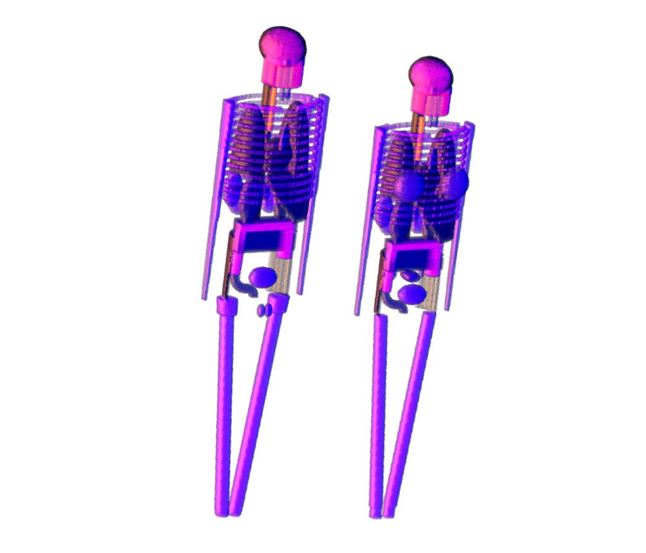
Figure 1
Views of ADAM and EVA.
In the voxelization of the pair, the applications FANTOMAS and IDN (Images in Numerical Dosimetry) were used, which was also developed in Windows C API, mainly for reading and writing binary files containing the 3D matrix corresponding to a stack of transverse images of a given geometry [24]. Among other skills, the IDN uses some CT image segmentation methods [25] and creates voxel phantoms containing arrangements of geometric shapes such as parallelepipeds, spheres, and cylinders. Currently, phantom voxelization tasks are concentrated in the DIP.
To overcome the obvious anatomical limitations of mathematical phantoms, such as distance between organs, voxel phantoms were introduced by Gibbs and collaborators in 1984 [26] and, independently, by Williams and collaborators in 1986 [27]. During the 1980s and 1990s several voxel phantoms were published (see a good review in [1]). Between 1994 and 1995, Zubal and collaborators [28,29,30] segmented CT (Computed Tomography) and MRI (Magnetic Resonance Imaging) images of an adult male patient scanned from the head to the half thighs, which resulted in the publication of three voxel phantoms, currently available http://noodle.med.yale.edu/zubal/: VOXEL_MAN, MAN_TISSUE3-6 and VOX_TISSUE8. Figure 2 shows views of these phantoms.
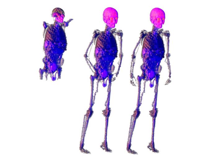
Figure 2
Views of the VOXEL_MAN, MAN_TISSUE3-6 and VOX_TISSUE8 phantoms.
In 2003, the ICRP published its report number 89 under the title, Basic Anatomical and Physiological Data for Use in Radiological Protection: Reference Values. This dataset was one of the motivations for the development of the MAX phantom. MAX was "born" from the VOXTISS8 voxel phantom, which represents an adult male available in a RAW file called vox_tiss8.dat, containing 85 segmented organs/tissues. These primary data were resampled, reclassified, and adjusted based on the ICRP 89 reference man. The detailed description of the resulting phantom and the new methods to estimate the equivalent dose in red bone marrow (RBM) and in the skin were topics of Vieira's doctoral thesis [1]. In the same year, Kramer and collaborators presented the FAX phantom [7], built from CT images based on anatomical data from ICRP 89.
FAX and MAX were resampled generating FAX06 and MAX06 [31], where the micro-CT method was implemented [32,33]. The 2006 article is cited in ICRP 110 and the 2010A one was published as one of the chapters of the book edited by Xu and Eckerman, which synthesizes the golden age of voxel phantoms. Figure 3 shows views of the FAX, MAX, FAX06 and MAX06 phantoms. With the resampling was possible to solve the skin dosimetry problem and improved the red bone marrow dosimetry method.
In 2009, with the purpose of standardizing the adult voxel phantoms and updating the CCs for estimating the dose in organs and tissues in radiological protection, the ICRP made available the ICRP-AM (ICRP Adult Male) and the ICRP-AF (ICRP Adult Female) in its report nº 110.
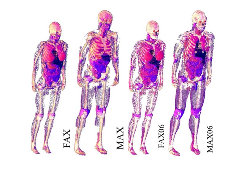
Figure 3
Views of the FAX, MAX, FAX06 and MAX06 phantoms.
3. MESH PHANTOMS AND VOXELIZATION
One of the main difficulties in producing voxel phantoms is the availability of a complete set of images of the human body. Thus, synthetic phantoms [16] emerged, that is, produced on the computer from 3D objects representing organs and tissues of the human body, obtained from specialized pages on the Internet or drawn based on information contained in anatomy books. In applications that edit polygonal meshes, 3D objects are assembled and adjusted, resulting in phantoms that are subsequently voxelized [34] for coupling to an MC code, forming an ECM.
For GDN, the most representative synthetic phantoms up to the present moment are MASH (Male Adult meSH) and FASH (Female Adult meSH), used in several significant publications of the group. For example, the article by Cassola and collaborators (2010) [35] is one of the references in ICRP report 145 on mesh phantoms. In 2011, using Blender, MakeHuman and ImageJ applications, Cassola built several mesh phantoms varying in posture, mass, and height [36]. Figure 4 represents views of the standing FASH and MASH phantoms, and their supine versions, FASH_SUP and MASH_SUP. These basic phantoms have total mass, organ masses and height in accordance with ICRP 89 [9] data. The four MCEs resulting from coupling them to EGSnrc, named FSTA, FSUP, MSTA and MSUP, are available at the DEN-UFPE page (http://www.caldose.org/). The twelve algorithms of radioactive sources for dosimetry (eleven for external and one for internal) [1], whose labels and names are listed in Table 1, complete these ECMs.
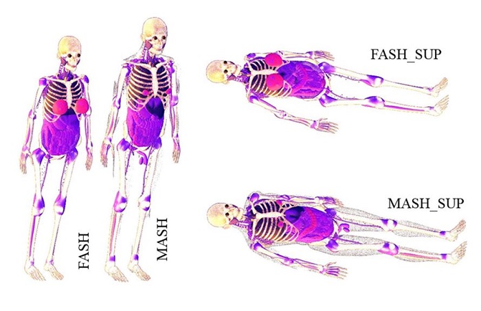
Figure 4
Views of FASH, MASH, FASH_SUP and MASH_SUP phantoms.
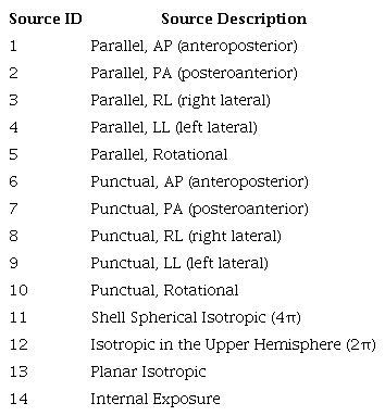
Also in 2011, applications such as MakeHuman, Blender, ImageJ and DIP were used to develop pediatric mesh phantoms, morphologically consistent with human anatomy, for use in dosimetry [37,38]. The two couple of children (5 and 10 years old) produced in these works were voxelized and will be part of a library of phantoms that the GDN is organizing for dosimetric evaluations. Figure 5 are views of them.
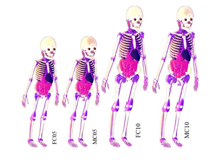
Figure 5
GDN pediatric phantoms.
In 2015, Cabral developed the MARIA mesh phantom (Anthropomorphic Model for the dosimetry of Ionizing Radiation in Adults), using the Autodesk 3ds Max application, from 3D objects obtained on the internet for the anatomical representation of a non-pregnant adult. The changes to 3D objects for the final representation of a pregnant woman were also made in 3ds Max [39].
In 2016, Santos developed the SARA mesh phantom (Anthropomorphic Simulator for Dosimetry of Ionizing Radiation in Adolescents) [40] and used the voxelized version [41] to simulate a craniospinal radiotherapy treatment, representing the LINAC (Linear Accelerator) from a phase space produced in the Quimera application [42]. The phase space consisted of a text file with information about the initial state of the photons, mainly the position, flight direction and energy, simulated data at an informed distance from the phantom and according to the area you want to radiate.
The male adult phantom MARTIN (Male Adult with Macro Circulation and Lymphatic Vessels Phantom) [43] and male adolescent SAMUEL (Male Anthropomorphic Simulator for Ionizing Radiation Dosimetry in Adolescents) [44] complete the anthropomorphic models revisited in this work. Figure 6 comprises views of the SARA, SAMUEL, MARIA, and MARTIN phantoms.
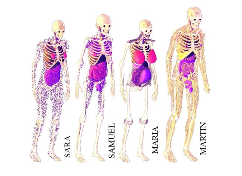
Figure 6
GDN teen and adult phantoms.
4. CONCLUSIONS AND PERSPECTIVES
In this article, a review of anthropomorphic computational phantoms used for simulations in internal and external dosimetry was carried out. These phantoms were produced or transformed in the three dissertations [39,40,44] and four theses [1,37,38,43] developed at DEN-UFPE, between 2004 and 2021. To produce phantoms, their coupling to MC codes and obtaining different dosimetric results, the GDN developed computational tools, such as the DIP application, for assembling MCEs using a voxel phantom and EGSnrc.
From voxelized versions of mathematical phantoms to mesh phantoms, there has been a significant improvement in realism and accuracy in modeling these 3D geometries by GDN. Figure 7 gives an idea of how the phantoms have been improved. By observing the shapes of some radiosensitive organs and bones in the three views of the Figure 7, it can be concluded that, under the same irradiation conditions, the numerical dosimetry performed with MARTIN will be the most accurate.
Figure 7
Views of some bones and radiosensitive organs of the ADAM, MAX and MARTIN phantoms.
At present the GDN needs to voxelize the mesh phantoms to couple them to the MC codes and perform dosimetric evaluations. The problem of coupling a mesh phantom directly to an MC code is still a challenge for the group, although it has already been carried out by other groups. Thus, the future production of dosimetric data with the couples whose views appear in Figures 5 and 6 will be the next step for the group and, in principle, with their voxelized versions.
ACKNOWLEDGMENT
The authors are grateful the Conselho Nacional de Desenvolvimento Científico e Tecnológico (CNPq), the Fundação de Amparo a Ciência e Tecnologia de Pernambuco (FACEPE) and the Instituto Federal de Pernambuco (IFPE) - Campus Recife for the financial support of and the Centro Regional de Ciências Nucleares do Nordeste (CRCN-NE) for the computational infrastructure that allowed us to perform the work included in this article.
REFERENCES
VIEIRA, J. W. Construção de um Modelo Computacional de Exposição para Cálculos Do-simétricos Utilizando o Código Monte Carlo EGS4 e Fantomas de Voxels, Tese de Doutora-do, Departamento de Energia Nuclear, Universidade Federal de Pernambuco, Recife, Pernam-buco, Brasil, 2004.
VIEIRA, J. W.; STOSIC, B.; LIMA, F. R. A.; KRAMER, R.; SANTOS, A. M.; LIMA, V. J. M. Um Aplicativo para Editar Fantomas de Voxels e Calcular Coeficientes de Conversão para a Proteção Radiológica. In: INTERNATIONAL JOINT CONFERENCE RADIO 2005, Annals RADIO 2005, ABENDE, Rio de Janeiro, 2005.
VIEIRA, J. W.; LIMA, F. R. A. A Software to Digital Image Processing to Be Used in the Voxel Phantom Development, Cell. Mol. Biol., v. 55, n. 3, p. 16-22, 2009.
PETZOLD, C. Programming Windows, Microsoft Press, USA, 1999
NELSON, W. R.; HIRAYAMA, H.; ROGERS, D. W. O. The EGS4 Code System, Report SLAC-265, Stanford Linear Accelerator Center, Stanford University, Stanford, USA, 1985.
KRAMER, R.; VIEIRA, J. W.; KHOURY, H. J.; LIMA, F. R. A.; FUELLE. D. All about MAX: A Male Adult Voxel Phantom for Monte Carlo Calculations in the Area of Radiation Protection Dosimetry, Phys. Med. Biol., v. 48, p. 1239-1262, 2003.
KRAMER, R.; KHOURY, H. J.; VIEIRA, J. W.; LOUREIRO, E. C. M.; LIMA, V. J. M.; LI-MA, F. R. A.; HOFF, G. All About Fax: A Female Adult Voxel Phantom for Monte Carlo Cal-culation in Radiation Protection Dosimetry, Phys. Med. Biol., v. 49, p. 5203-5216, 2004A
ICRP 60, 1990 Recommendations of the International Commission on Radiological Protec-tion, ICRP Publication 60, Ann. ICRP 21 (1-3), 1991.
ICRP 89, Basic Anatomical and Physiological Data for Use in Radiological Protection: Ref-erence Values, ICRP Publication 89, Pergamon Press, Oxford, 2003.
TEMPLEMAN, J.; OLSEN, A. Microsoft Visual C++ .NET, Step by Step, Microsoft Press, USA, 2002.
SHARP, J. Microsoft Visual C# 2013: Passo a Passo, Bookman, Porto Alegre, RS, Brasil, 2014.
VIEIRA, J. W.; LEAL NETO, V.; LIMA FILHO, J. M.; LIMA, F. R. A. Desenvolvimento de Algoritmos Simuladores de Fontes Radioativas Planares para Uso em Modelos Computacionais de Exposição, Brazilian Journal of Radiation Sciences, v. 1, p. 1-17, 2013.
KAWRAKOW, I.; MAINEGRA-HING, E.; ROGERS, D. W. O.; TESSIER, F.; WALTERS, B. R. B. The EGSnrc Code System: Monte Carlo simulation of electron and photon transport. Technical Report PIRS-701. National Research Council Canada, Ottawa, 2021.
VIEIRA, J. W. MonteCarlo – Um Aplicativo para Uso em Avaliações Dosimétricas das Radiações Ionizantes, Tese para Progressão à Classe Titular do IFPE, Recife, Pernambuco, Brasil, 2017.
ICRP 145, Adult Mesh-Type Reference Computational Phantoms, ICRP Publication 145, ICRP 49(3), 2020.
GONZALEZ, R. C.; WOODS, R. E. Processamento Digital de Imagens, Pearson Education do Brasil, São Paulo, SP, Brasil, 2010.
FISHER. H. L.; SNYDER, W. S. Distribution of Dose in the Body from a Source of Gam-ma Rays Distributed Uniformly in an Organ, Report n. ORNL-4168, Oak Ridge National Laboratory, Oak Ridge, Tenn., USA, 1967.
ICRP 23, Report of the Task Group on Reference Man, International Commission on Radi-ological Protection, Pergamon Press, Oxford, 1975.
SNYDER, W. S.; FORD, M. R.; WARNER, G. G.; FISHER, H. L. Estimates of Absorbed Fractions for Monoenergetic Photon Sources Uniformly Distributed in Various Organs of a Het-erogeneous Phantom, MIRD Pamphlet n. 5, J. Nucl. Med., v. 10: Suppl. n. 3, p. 7-52, 1969.
KRAMER, R.; ZANKL, M.; WILLIAMS, G.; DREXLER, G. The Calculation of Dose from External Photon Exposures Using Reference Human Phantoms and Monte Carlo Methods. Part I: The Male (ADAM) and Female (EVA) Adult Mathematical Phantoms, GSF-Bericht S-885, GSF-National Research for Environment and Health, Neuherberg, 1982.
KRAMER, R.; VIEIRA, J. W.; KHOURY, H. J.; LIMA, F. R. A. MAX Meets ADAM: A Do-simetric Comparison between a Voxel-Based and a Mathematical Model for External Exposure to Photons, Phys. Med. Biol., v. 49, p. 887-910, 2004B.
ICRP 110, Adult Reference Computational Phantoms, ICRP Publication 110, Elsevier Ltd, 2009.
ICRP 147, Use of Dose Quantities in Radiological Protection, ICRP Publication 147, Ann. ICRP 50(1), 2021.
VIEIRA, J. W.; SANTOS, A. M.; LIMA, F. R. A. Tratamento de Imagens Tomográficas para Uso em Dosimetria Numérica, In: First American IRPA Congress, XXIV SMSR Annual Meeting XVII Annual SNM Congress, Acapulco, 2006.
NIKOLAIDIS, N. PITAS, I. 3-D Image Processing Algorithms, USA: John Wiley Sons, 2001.
GIBBS, S. J.; PUJOL, A.; CHEN, T-S.; MALCOLM, A. W.; JAMES, A. E. Patient Risk from Interproximal Radiography, Oral Surg. Oral Med. Oral Pathol., v. 58, p. 347-354, 1984.
WILLIAMS, G.; ZANKL, M.; ABMAYR, W.; VEIT, R.; DREXLER, G. The Calculation of Dose from External Photon Exposures Using Reference and Realistic Human Phantoms and Monte Carlo Methods, Phys. Med. Biol., v. 31, p. 347-354, 1986.
ZUBAL, I. G.; HARRELL, C. R.; SMITH, E. O.; RATTNER, Z.; GINDI, G.; HOFFER, P. B. Computerized Three-Dimensional Segmented Human Anatomy, Med. Phys., v. 21 (2), p. 299-302, 1994a.
ZUBAL, I. G.; HARRELL, C. R.; SMITH, E. O.; SMITH, A. L.; KRISCHLUNAS, P. High Resolution, MRI-Based, Segmented, Computerized Head Phantom, 1994b. Available at http://noodle.med.yale.edu/zubal>.
ZUBAL, I. G.; HARRELL, C. R.; SMITH, E. O.; SMITH, A. L. Two Dedicated Software, Voxel-Based, Anthropomorphic (Torso and Head) Phantoms, In: Proceedings of an Interna-tional Workshop on Voxel Phantom Development held at the National Radiological Protection Board, Chilton, UK, 6-7 July, 1995.
KRAMER, R.; KHOURY, H. J.; VIEIRA, J. W.; LIMA, V. J. M. MAX06 and FAX06: Update of Two Adult Human Phantoms for Radiation Protection Dosimetry, Phys. Med. Biol., v. 51, p. 3331-3346, 2006.
KRAMER, R.; KHOURY, H. J.; VIEIRA, J. W.; KAWRAKOW, I. Skeletal Dosimetry for External Exposure to Photons Based on μCT Images of Spongiosa from Different Bone Sites, Phys. Med. Biol., v. 52, p. 6697-6716, 2007.
KRAMER, R.; KHOURY, H. J.; VIEIRA, J. W.; LIMA, V. J. M.; LOUREIRO, E. C. M.; HOFF, G.; KAWRAKOW, I. The FAX06 and the MAX06 Computational Voxel Phantoms, In Xu XG, Eckerman KF (eds.), Handbook of Anatomical Models for Radiation Dosimetry, 2010A, (Series in Medical Physics and Biomedical Engineering, CRC Press).
ANDRADE, P. H. A.; VIEIRA, J. W.; OLIVEIRA, V. R. S.; VELOSO, R. J. B.; LIMA, F. R. A. Um Método para Voxelização de Geometrias 3D de Malhas, Brazilian Journal of Radia-tion Sciences, v. 08-01A, p. 01-10, 2020.
CASSOLA, V. F.; LIMA, V. J. M.; KRAMER, R.; KHOURY, H. J.; FASH and MASH: Fe-male and Male Adult Human Phantoms Based on Polygon Mesh Surfaces: I. Development of the Anatomy, Phys. Med. Biol., v. 55, p. 133-162, 2010.
CASSOLA, V. F. Desenvolvimento de Fantomas Humanos Computacionais Usando Ma-lhas Poligonais em Função da Postura, Massa e Altura, Tese de Doutorado, Departamento de Energia Nuclear, Universidade Federal de Pernambuco, Recife, Pernambuco, Brasil, 2011.
LIMA, V. J. M. Desenvolvimento de Fantomas MESH Infantis, Morfologicamente Consis-tentes com a Anatomia Humana, para Uso em Dosimetria, Tese de Doutorado, Departamento de Energia Nuclear, Universidade Federal de Pernambuco, Recife, Pernambuco, Brasil, 2011.
LIMA, V. J. M.; CASSOLA, V. F.; KRAMER, R.; LIRA, C. A. B. O.; KHOURY, H. J. De-velopment of 5- and 10-Year-Old Pediatric Phantoms Based on Polygon MESH Surfaces, Med. Phys., v. 38 (8), 2011.
CABRAL, M. O. M. Desenvolvimento de um Modelo Computacional de Exposição para Uso em Avaliações Dosimétricas em Gestantes, Dissertação de Mestrado, Departamento de Energia Nuclear, Universidade Federal de Pernambuco, Recife, Pernambuco, Brasil, 2015.
SANTOS, P. N. C. Simulação de um Tratamento Radioterápico Crânio-Espinhal em um Fantoma de Voxel Infantil Utilizando Espaços de Fase Representativos de um Acelerador Linear, Dissertação de Mestrado, Departamento de Energia Nuclear, Universidade Federal de Pernambuco, Recife, Pernambuco, Brasil, 2016.
VIEIRA, J. W.; CABRAL, M. O. M.; ANDRADE, P. H. A.; LEAL NETO, V.; LIMA, V. J. M.; LIMA FILHO, J. M.; LIMA. F. R. A. Uso do Software DIP para Voxelização de Fanto-mas MESH, In: V Congresso de Proteção Contra Radiações da Comunidade dos Países de Lín-gua Portuguesa, Coimbra, 2016.
OLIVEIRA, A. C. H. Desenvolvimento de um Sistema Computacional Baseado no Código Geant4 para Avaliações Dosimétricas em Radioterapia, Tese de Doutorado, Departamento de Energia Nuclear, Universidade Federal de Pernambuco, Recife, Pernambuco, Brasil, 2016.
ANDRADE, P. H. A. Construção e Voxelização de um Fantoma MESH Masculino Adulto com Macro Circulação e Vasos Linfáticos, Tese de Doutorado, Departamento de Energia Nu-clear, Universidade Federal de Pernambuco, Recife, Pernambuco, Brasil, 2018.
OLIVEIRA, E. S.; Estimativa da Dose Efetiva em Fantomas de Voxel para Indivíduos de 15 Anos, Dissertação de Mestrado, Departamento de Energia Nuclear, Universidade Federal de Pernambuco, Recife, Pernambuco, Brasil, 2021.
Notes
Author notes
*jose.wilson@recife.ifpe.edu.br
Conflict of interest declaration