Artículos
Abstract: Selenium (Se) is a trace element important for performance and immune functions in cattle. The deficiency of this mineral is widely distributed in northwestern Argentina and it is usually associated with intensive production systems. Calves seem more susceptible due to the metabolic regulation failures produced by this deficiency. This paper aims to describe a case of acute muscular dystrophy associated with Selenium deficiency in seven calves from a dairy farm in Salta, Argentina between March and May 2022. Selenium levels in the animals’ whole blood revealed that 66% of the calves (68,31±10,52 ppb) and approximately 90% of the cows (post-partum=71,24±10,04 ppb, and pre-partum 97,68±36,35 ppb) presented severely deficient values. The animals showed dehydration, weakness, prostration, respiratory signs, and even death without perceptible clinical signs. Macroscopically, diffuse pallor was found in the muscles of the hind limbs and in the myocardium in certain cases. Microscopically, severe polyphasic degeneration and necrosis of the muscle fibers were found, consistent with acute muscular dystrophy.
Keywords: nutritional muscular dystrophy, white muscle disease, perinatal mortality, deficiency disease, selenium.
Resumen: El selenio (Se) es un oligoelemento importante para que el ganado mantenga sus funciones inmunológicas adecuadamente y logre un correcto desempeño productivo. La deficiencia de este mineral está ampliamente distribuida en el noroeste de Argentina y suele estar asociada con sistemas de producción intensivos. Los animales jóvenes parecen ser más susceptibles debido a fallas en la regulación metabólica producidas por esta deficiencia. Este artículo tiene como objetivo describir un caso de distrofia muscular aguda asociada con la deficiencia de selenio en terneros de un establecimiento lechero ubicado en Salta, Argentina, entre marzo y mayo de 2022. Los niveles de selenio en la sangre total de los animales revelaron que el 66% de los terneros (68,31±10,52 ppb) y aproximadamente el 90% de las vacas (postparto = 71,24±10,04 ppb y preparto 97,68±36,35 ppb) presentaban valores de deficiencia severa. Los animales mostraban deshidratación, debilidad, postración, signos respiratorios o incluso muerte sin signos clínicos perceptibles. Macroscópicamente, se encontró una palidez difusa en los músculos de las extremidades posteriores y del miocardio en ciertos casos. Microscópicamente, se encontraron degeneración polifásica severa y necrosis de las fibras musculares, consistente con la distrofia muscular aguda.
Palabras clave: distrofia muscular nutricional, enfermedad de músculo blanco, mortalidad perinatal, enfermedad carencial, selenio.
INTRODUCTION
Selenium (Se) is a trace element that plays a significant role in the health and performance of cattle (Sapkota, 2020). The incidence of selenium deficiency is distributed in various regions worldwide and has become more important in recent years because of the intensification of the systems (Mehdi and Dufrasne, 2016). In cattle, this deficiency affects both performance (Hall et al., 2013b) and immune functions (Hall et al., 2013a; Hugejiletu et al., 2013).
Young animals are more susceptible to clinical disorders associated with selenium deficiency because the metabolic pathways in developing organisms are more easily deregulated than those in adult cattle (Hall et al., 2014). In newborn calves, weakness at birth and high perinatal mortality rates associated with secondary infections can be observed (Bostedt and Schramel, 1990). In cases where the deficiency is severe, cases of nutritional muscular dystrophy may occur (Ammerman and Miller, 1975). The transfer of selenium from cows to newborn calves is done through the placenta and milk (Enjalbert et al., 1999). For this reason, it is essential to ensure the correct supplementation of pregnant cows during the last third of pregnancy (Bayril et al., 2015).
This paper describes an acute case of muscular dystrophy associated with high mortality rates in dairy calves in an intensive dairy farm in northwestern Argentina. Clinical, pathological, epidemiological, and biochemical aspects are highlighted.
CASE DESCRIPTION
Between March and May 2022, the Specialized Veterinary Diagnostic Service from INTA Salta (SDVE-INTA Salta) provided diagnostic assistance to a dairy farm in the town of “Campo Quijano” in Salta province due to a gradual increase in the perinatal and neonatal mortality rates of calves in an artificial rearing system. In the mentioned period, fifteen deaths out of forty-five births were registered. The newborn animals were immediately separated from their mothers and taken to a calf barn where they were offered three liters of colostrum each (by voluntary intake or by tube). Injectable iodine was applied to them too. On average, protein determination in the serum of colostrated animals using a refractometer ranged from 6.8 to 7.1 mg/mL. Before admission, the floor of the rearing area was incinerated, and each calf was housed in an individual pen with bedding and shelter. A total of seven complete necropsies were performed, including sampling for microbiological and histopathological studies. Additionally, blood samples were collected from prepartum and postpartum females and newborn calves.
RESULTS
Clinical findings and epidemiological findings
The affected calves were less than 10 days old. The animals died within one to two days after the onset of signs; a moderate degree of dehydration, extreme weakness, prostration, respiratory signs, and vocalizations were observed. In four animals, death occurred without previous signs. Only three of the affected calves showed diarrhea. The monthly mortality data recorded are shown in fig. 1.
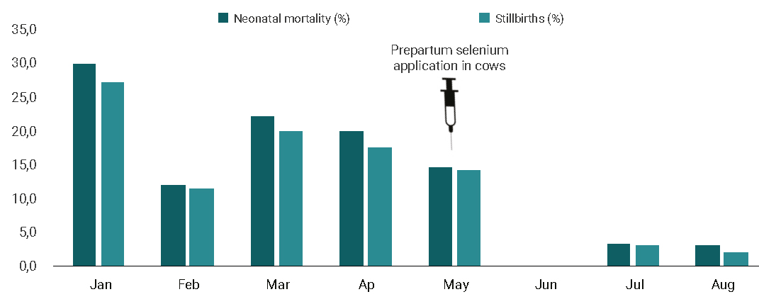
Figure 1
Epidemiological data. Neonatal mortality (%) and Stillbirths (%) rates in calves between the periods Jan-Aug 2022.
Microbiological findings
Lung, spleen and liver samples were taken and cultured from all necropsied animals. All samples were negative for relevant pathogens.
Pathological findings
Pathological findings consistent with muscular dystrophy were present in three of the seven dead calves (42%). In the remaining four calves, no lesions were detected in the muscles evaluated. The findings are shown in fig. 2. The macroscopic lesions were characterized by a diffuse pallor in extensive muscle areas, especially in the muscles of the hind limbs. The most severely affected muscles were the semimembranosus, semitendinosus, and quadriceps. In two animals, whitish lesions were observed at the level of the myocardium. A histopathology study revealed severe polyphasic degeneration and necrosis of the muscle fibers, consistent with acute muscular dystrophy. The muscle fibers were hyper-eosinophilic, fragmented, and vacuolated and showed loss of cross-striation with the influx of macrophages and hypertrophy of satellite cells. A small number of muscle fibers retained some striation, showed increased basophilia and centrally localized nuclei, suggesting regeneration.
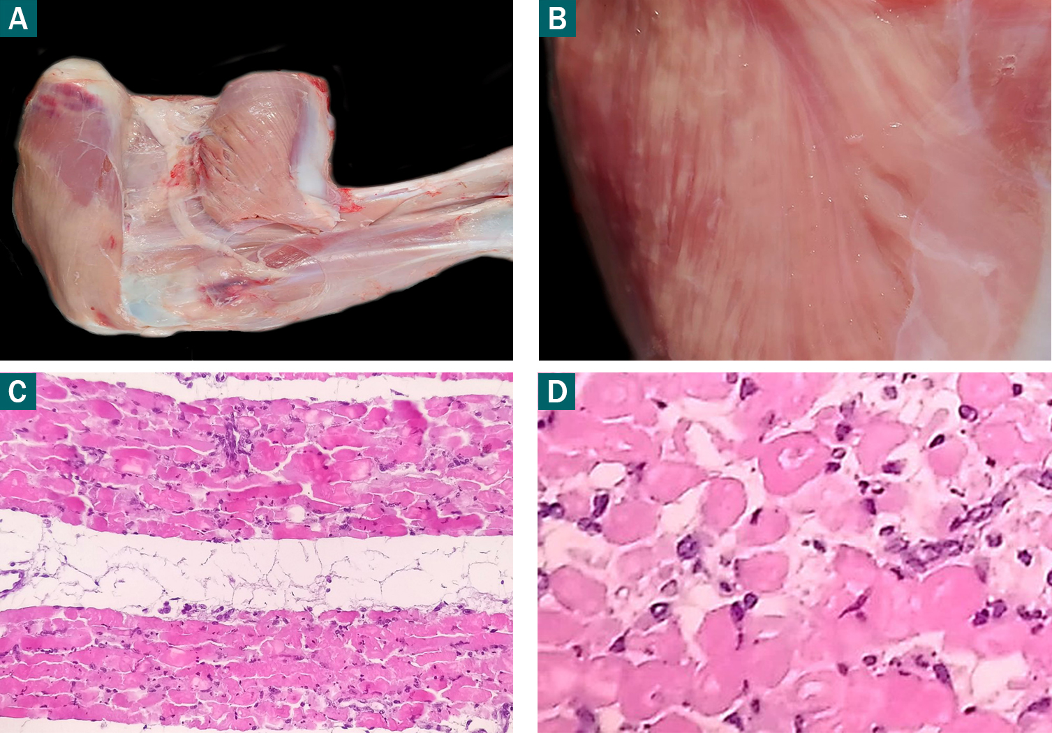
Figure 2
A. Macroscopic finding in muscular dystrophy due to Se deficiency in the limbs. Note the pale areas interspersed with normal areas. B. Muscular dystrophy in hind limbs muscle. C, D. Longitudinal and transverse cut sections of striated skeletal muscle, respectively. Hypereosinophilia, fragmentation, and vacuolization of the myofibrils (10X, H&E).
Biochemical findings
The concentrations of Se in the whole blood of prepartum and postpartum cows and newborn calves are shown in table 1. Briefly, low levels of selenium were observed in a high percentage of pre-partum (97,7±36,4 ppb) and post-partum (71,2±10,0 ppb) cows, and in newborn calves (68,3±10,5 ppb). In cattle, selenium values greater than 210 ppb are considered normal. In this case, the levels were much lower than those established in the bibliography (Kinkaid, 2000).
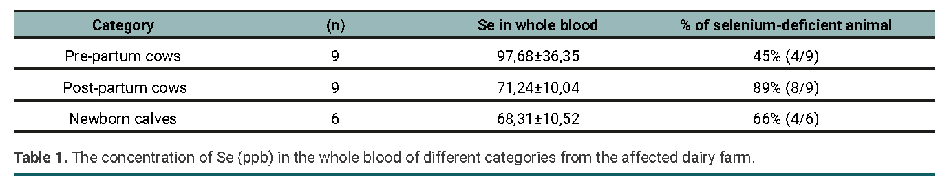
Table 1
The concentration of Se (ppb) in the whole blood of different categories from the affected dairy farm.
DISCUSSION AND CONCLUSIONS
Nutritional muscular dystrophy (NMD), or “white muscle disease”, is a pathological entity associated with severe Se and/or vitamin E deficiency (Andersonet al., 1977; Maas, 1984). In the present work, the clinical and pathological findings associated with the low levels of Se detected in calves and cows suggest that the deficiency of this mineral was the trigger for the high mortality rates observed. It should be noted that NMD is a late expression of selenium deficiency (Mehdi and Dufrasne, 2016). Other diseases can cause similar muscle injuries, including poisoning by ingesting Senna occidentalis (Carmo et al., 2011) or ionophore toxicity, such as monensin poisoning (Brito et al., 2016). Those causes were ruled out based on the anamnestic and epidemiological data. On the other hand, no studies could be conducted on blood tocopherol levels. Because of this, the possibility of a combined deficiency of Se and vitamin E could not be excluded. The response to the treatment with Se reaffirmed the origin of the disease.
The severity of the pathological changes produced by selenium deficiency depends on age, which also determines whether the consequences are fatal or not (Sapkota, 2020). Pathological conditions and deaths owing to selenium deficiency occur primarily in newborn animals and those in the early and last postnatal stages of development. The reasons why selenium deficiency results in such a diverse range of unusual pathological conditions are still unclear (Mehdi and Dufrasne, 2016). Because vitamin E and selenium have interdependent antioxidant functions, a deficiency of one of these compounds may increase the requirements of the other needed to prevent abnormalities (NRC 2000). It is likely that the cause of selenium-deficient animals manifesting one or any of the other symptoms depends on the concomitant vitamin E deficiency (Sapkota, 2020).
Kinkaid (2000) mentions that values of selenium in blood below 60 ppb indicate “severe deficiency”, values between 60 and 200 ppb are “marginally deficient”, values between 210 and 1200 ppb are “adequate” and greater than 1200 ppb are highly adequate. In this report, the results of selenium in the whole blood obtained from cows and calves showed low levels of this micro-mineral. About 66% of the calves and approximately 90% of the cows showed severe Se deficiency.
In Argentina, cases of nutritional muscular dystrophy have been registered in cattle (Rodriguez et al., 2018) and sheep (Micheloud et al., 2018), although information about that disease in dairy herds is scarce. However, selenium deficiency is widespread throughout the country (Lizárraga, 2021). In this regard, in a study conducted by Cseh et al. (2013) in which the level of glutathione peroxidase (GPx) was determined in 3621 blood samples of beef cattle from different farms in Argentina, it was found that 36% of the animals presented values below 30 IU/g of hemoglobin, which were compatible with Se deficiency.
Another author (Minatel et al., 2004) conducted a study in the northwestern region of Buenos Aires province, Argentina, and observed that 77% and 68.5% of the beef cattle blood samples presented compatible levels with deficiency of Se during winter and summer, respectively.
Several studies have suggested an immunosuppressive effect of selenium deficiency in ruminants (MacPherson, 1994). For example, this deficiency has been associated with sepsis in young cattle and mastitis in adult cows (Dalir-Naghadeh et al., 2006). It has been suggested that marginal selenium deficiency may be associated with general morbidity and/or poor performance in calves (Waltner-Toews et al., 1986). Consistent with this, Spears et al. (1986) show a lower incidence of neonatal diarrhea and a reduction in mortality in beef calves supplemented with this mineral, and how stillbirths and neonatal mortality were significantly reduced following the application of injectable selenium to prepartum cows.
Finally, due to the productive and reproductive importance of this micromineral, selenium deficiency must be considered when addressing morbidity or mortality issues in artificially fed calves.
REFERENCES
ANDERSON, P.H.; BRADLEY, R.; BERRETT, S.; PATTERSON, D.S.P. 1977. The sequence of myodegeneration in nutritional myopathy of the older calf. The British Veterinary Journal 133, 160-165.
AMMERMAN, C.B.; MILLER, S.M. 1975. Selenium in ruminant nutrition: a review. Journal of Dairy Science 58, 1561-1577.
BAYRIL, T.; YILDIZ, A.S.; AKDEMIR, F.; YALCIN, C.; KÖSE, M.; YILMAZ, O. 2015. The Technical and Financial Effects of Parenteral Supplementation with Selenium and Vitamin E during Late Pregnancy and the Early Lactation Period on the Productivity of Dairy Cattle. Asian-Australas J Anim Sci. 28(8):1133-1139. https://doi.org/10.5713/ajas.14.0960
BOSTEDT, H.; SCHRAMEL, P. 1990. The importance of selenium in the prenatal and postnatal development of calves and lambs. Biological trace element research, 24(2), 163-171. https://doi.org/10.1007/BF02917204
BRITO, E.S.A.; ANDRADE, T.G.; OLIVEIRA, C.H.S.D.; MOURA, V.M.B.D.D. 2020. The outbreak of monensin poisoning in cattle due to supplementation error. Ciencia Rural, 50 (11). https://doi.org/10.1590/0103-8478cr20190996
CARMO, P.; IRIGOYEN, L.F.; LUCENA, R.B.; FIGHERA, R.A.; KOMMERS, G.D.; BARROS, C.S. 2011. Spontaneous coffee senna poisoning in cattle: report on 16 outbreaks. Pesquisa Veterinária Brasileira, 31(2), 139-146. https://doi.org/10.1590/S0100-736X2011000200008
CSEH, S.B.; DRAKE, M.L.; BRAMBILLA, E.C. 2013. Deficiencia de selenio en bovinos según la época del año y región en Argentina. Revista de Producción Animal, 33(3):21.
DALIR-NAGHADEH, B.; ASRI-REZAEI, S.; ALIDADY, N. 2006. Measure of association between selenium status and sepsis in cattle. Comp Clin Pathol 15, 149-153. https://doi.org/10.1007/s00580-006-0622-6
ENJALBERT, F.; LEBRETON, P.; SALAT, O.; SCHELCHER, F. 1999. Effects of pre- or postpartum selenium supplementation on selenium status in beef cows and their calves. J. Anim. Sci., 77 (1), 223-229. https://doi.org/10.2527/1999.771223x
HALL, J.A.; BOBE, G.; VORACHEK, W.R.; ESTILL, C.T.; MOSHER, W.D.; PIRELLI, G.J.; GAMROTH, M. 2014. Effect of supranutritional maternal and colostral selenium supplementation on passive absorption of immunoglobulin G in selenium-replete dairy calves. Journal of Dairy Science, 97(7), 4379-4391. https://doi.org/10.3168/jds.2013-7481
HALL, J.A.; BOBE, G.; HUNTER, J.K.; VORACHEK, W.R.; STEWART, W.C.; VANEGAS, J.A.; ESTILL, C.T.; MOSHER, W.D.; PIRELLI, G.J. 2013a. Effect of feeding selenium-fertilized alfalfa hay on performance of weaned beef calves. PLoS ONE 8:e5818.
HALL, J.A.; BOBE, G.; VORACHEK, W.R.; HUGEJILETU; GORMAN, M.E.; MOSHER, W.D.; PIRELLI, G.J. 2013b. Effects of feeding selenium-enriched alfalfa hay on immunity and health of weaned beef calves. Biol. Trace Elem. Res. 156:96-110.
HUGEJILETU; BOBE, G.; VORACHEK, W.R.; GORMAN, M.E.; MOSHER, W.D.; PIRELLI, G.J.; HALL, J.A. 2013. Selenium supplementation alters gene expression profiles associated with innate immunity in whole-blood neutrophils of sheep. Biol. Trace Elem. Res. 154:28-44.
KINCAID, R.L. 2000. Assessment of trace mineral status of ruminants: a review. J. Anim. Sci. 77, 1-10.
LIZÁRRAGA, R.M. 2021. Consecuencias productivas y reproductivas de la deficiencia de selenio en bovinos. Tesis doctoral, Universidad Nacional de La Plata. oai:sedici.unlp.edu.ar:10915/126267
MAAS, J.; BULGIN, M.S.; ANDERSON, B.C.; FRYE, T.M. 1984. Nutritional myodegeneration associated with vitamin E deficiency and normal selenium status in lambs. Journal of the American Veterinary Medical Association 184, 201-204.
MACPHERSON, A. 1994. Selenium, vitamin E biological oxidation. In: GARNSWORTHY, P.C.; COLE, D.J.A. (eds.). Recent advances in animal nutrition. Nottingham University Press, Nottingham. 3-30 pp.
MEHDI, Y.; DUFRASNE, I. 2016. “Selenium in cattle: a review.” Molecules 21.4: 545.
MICHELOUD, J.F.; ARAOZ, V.; DELGADO, F.O.; COLQUE-CARO, L.A.; ROSA, D.E.; MATTIOLI, G.A. 2018. Distrofia muscular nutricional en corderos de raza Dorper en el Noroeste Argentino. Rev. Med. Vet. (Buenos Aires) 99(2): 13-16.
MINATEL, L.; BUFFARINI, M.A.; SCARLATA, E.F.; DALLORSO, M.E.; CARFAGNINI, J.C. 2004. Niveles de cobre, hierro, zinc y selenio de bovinos del noroeste de la provincia de Buenos Aires. Rev. Arg. Prod. Anim, 24(3-4), 225-235.
NATIONAL RESEARCH COUNCIL. 2000. Nutrient Requirements of Beef Cattle. Seventh Revised Edition; National Academy Press: Washington, DC, USA, 1996.
RODRIGUEZ, A.M.; SCHILD, C.O.; CANTÓN, G.J.; RIET-CORREA, F.; ARMENDANO, J.I.; CAFFARENA, R.D.; GIANNITTI, F. 2018. White muscle disease in three selenium deficient beef and dairy calves in Argentina and Uruguay. Ciência Rural, 48.
SAPKOTA, D. 2020. Selenium and vitamin E deficiency in animals. The Blue Cross, 16, 75-79.
SPEARS, J.W.; HARVEY, R.W.; SEGERSON, E.C. 1986. Effects of marginal selenium deficiency and winter protein suplementaction on growth, reproduction and selenium status of beef cattle. Journal animal science 67. 586-594.
WALTNER-TOEWS, D.; MARTIN, S.W.; MEEK, A.H. 1986. Selenium content in the hair of newborn dairy heifer calves and its association with preweaning morbidity and mortality. Canadian Journal of Veterinary Research, 50(3), 347.
WEISS, W.P.; COLENBRANDER, V.F.; CUNNINGHAM, M.D.; CALLAHAN, C.J. 1983. Selenium/vitamin E: role in disease prevention and weight gain of neonatal calves. Journal of dairy science 66.5 1101-1107.
